Review
. 2009:87:53-79.
doi: 10.1016/S0070-2153(09)01202-2.

Synapse formation in developing neural circuits

Affiliations
Review

Synapse formation in developing neural circuits

Daniel A Colón-Ramos. Curr Top Dev Biol. 2009.

Abstract

The nervous system consists of hundreds of billions of neurons interconnected into the functional neural networks that underlie behaviors. The capacity of a neuron to innervate and function within a network is mediated via specialized cell junctions known as synapses. Synapses are macromolecular structures that regulate intercellular communication in the nervous system, and are the main gatekeepers of information flow within neural networks. Where and when synapses form determines the connectivity and functionality of neural networks. Therefore, our knowledge of how synapse formation is regulated is critical to our understanding of the nervous system and how it goes awry in neurological disorders. Synapse formation involves pairing of the pre- and postsynaptic partners at a specific neurospatial coordinate. The specificity of synapse formation requires the precise execution of multiple developmental events, including cell fate specification, cell migration, axon guidance, dendritic growth, synaptic target selection, and synaptogenesis (Juttner and Rathjen in Cell. Mol. Life Sci. 62:2811, 2005; Salie et al., in Neuron 45:189, 2005; Waites et al., in Annu. Rev. Neurosci. 28:251, 2005). Remarkably, during the development of the vertebrate nervous system, these developmental processes occur almost simultaneously in billions of neurons, resulting in the formation of trillions of synapses. How this remarkable specificity is orchestrated during development is one of the outstanding questions in the field of neurobiology, and the focus of discussion of this chapter. We center the discussion of this chapter on the early developmental events that orchestrate the process of synaptogenesis prior to activity-dependent mechanisms. We have therefore limited the discussion of important activity-dependent synaptogenic events, which are discussed in other chapters of this book. Moreover, our discussion is biased toward lessons we have learned from invertebrate systems, in particular from C. elegans and Drosophila. We did so to complement the discussions from other chapters in this book, which focus on the important findings that have recently emerged from the vertebrate literature. The chapter begins with a brief history of the field of synaptic biology. This serves as a backdrop to introduce some of the historically outstanding questions of synaptic development that have eluded us during the past century, and which are the focus of this review. We then discuss some general features of synaptic structure as it relates to its function. In particular, we will highlight evolutionarily conserved traits shared by all synaptic structures, and how these features have helped optimize these ancient cellular junctions for interneural communication. We then discuss the regulatory signals that orchestrate the precise assembly of these conserved macromolecular structures. This discussion will be framed in the context of the neurodevelopmental process. Specifically, much of our discussion will focus on how the seemingly disparate developmental processes are intimately linked at a molecular level, and how this relationship might be crucial in the developmental orchestration of circuit assembly. We hope that the discussion of the multifunctional cues that direct circuit development provides a conceptual framework into understanding how, with a limited set of signaling molecules, precise neural wiring can be coordinated between synaptic partners.


Figures

Figure 2.1
Figure 2.1
Transcription factor Engrailed directs synaptic specificity in the cockroach cercal system. Wiring diagram of the cockroach cercal system in the second instar cockroach. (A) In wild-type animals, medial sensory neuron (neuron 6m, in yellow) expresses Engrailed (represented by dark nuclei), while lateral sensory neuron (6d, in blue) does not (represented by clear nuclei). Although the axonal arbors of the medial and lateral sensory neurons overlap, they display specificity by connecting with specific target interneurons: medial sensory neuron connects to interneuron G12 (in orange), while lateral sensory neuron connects to interneuron G13 (in green). This specificity can be physiologically recorded, so that, for instance, stimuli in the filiform hair linked to the medial sensory neuron (dark arrow) results in depolarization of interneuron G12 and not G13 (represented by physiological recording to the right of the schematic). (B) Loss of Engrailed by dsRNA disrupts the medial sensory neuron identity and connectivity. In the absence of Engrailed, the medial sensory neuron (6m) adopts an identity similar to the lateral sensory neuron in terms of the branching structure (compare branching schematic of 6m in A (yellow) with 6m in B (blue)). Loss of Engrailed also disrupts medial sensory neuron synaptic specificity (represented by physiological recording to the right of the schematic).
Figure 2.2
Figure 2.2
Axon guidance molecule Netrin is required for synaptic targeting events in the Drosophila embryo. Schematic diagram of RP3 neuron (blue) and the body wall muscles in the Drosophila embryo (represented here are muscles 6, 7, 13, 12, 5, and 8). (A) In wild-type animals, muscles 6 and 7 express Netrin (in pink). Muscles 12, 13, 5, and 8 express repulsive cues such as semaphorins, and do not express Netrin (lack of Netrin expression represented in white). Expression of Netrin by muscles 6 and 7 induces short-range targeting and specific innervation of these muscles by the RP3 neuron. (B) In Netrin loss-of-function mutants, the RP3 neuron reaches the neighborhood of muscles 6 and 7 in a timely fashion, coming within filopodial reach of its targets, but fails to innervate these muscles correctly. These studies suggest that Netrin is not required for long-range guidance of the RP3 neuron, but is instead required for its short-range synaptic targeting.
Figure 2.3
Figure 2.3
Guidepost cells direct synaptic specification. Schematic representing examples of guidepost cells directing synaptic specification in C. elegans. (A) Image of C. elegans with the discussed regions boxed. (B) Box-I: In the nematode head, interneuron AIY (black) contacts many neurons, but connects specifically to interneuron RIA (not shown) at a subcellular region of its neurite (boxed). This specificity is directed by ventral cephalic sheath cells (pink). Ventral cephalic sheath cells are glial cells that project a process posteriorly, where it contacts AIY and RIA, and also express Netrin. The expression of Netrin directs presynaptic assembly (in green) in the correct subcellular region of AIY (boxed region). Box-II: In the nematode egg-laying circuit, neuron HSNL (in black) innervates other neurons and muscles (not shown) in a specific and stereotyped fashion. The postsynaptic partners of HSNL are not required for the precise assembly of presynaptic specializations (in green) in a subcellular region of HSNL. Instead, guidepost epithelial cells (dark spheres) express an immunoglobulin superfamily receptor (SYG-2) that directs where synapses form in HSNL. Box-Ill: In the posterior part of the nematode, neuron DA9 (in black) elaborates a dendrite anteriorly within the ventral nerve cord and extends an axon commissurally and then longitudinally along the dorsal nerve cord. UNC-6/Netrin (pink) and LIN-44/Wnt (blue) direct synaptic specification (green) by inhibiting formation of synapses from discrete subcellular domains. Expression of lin-44/wnt (blue) by cells in the posterior part of the animal prevents ectopic synapse formation in the commissure, while expression of Netrin (pink) by ventral cells excludes presynaptic components from the ventral dendrite.
Figure 2.4
Figure 2.4
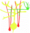
Bergmann glia direct the innervation of Stellate axons to the Purkinje dendrites. Purkinje neurons (yellow) are innervated by stellate interneurons (green) exclusively at the dendrites. This precision is directed by Bergmann glia (red), which act as guideposts by directing the stellate interneuron process to their Purkinje neuron targets (synapses in blue).

References

    1. Aberle H, Haghighi AP, Fetter RD, McCabe BD, Magalhaes TR, and Goodman CS (2002). Wishful thinking encodes a BMP type II receptor that regulates synaptic growth in Drosophila. Neuron 33, 545–558. - PubMed
    1. Ackley BD, Harrington RJ, Hudson ML, Williams L, Kenyon CJ, Chisholin AD, and Jin Y (2005). The two isoforms of the Caenorhabditis elegans leukocyte-common antigen related receptor tyrosine phosphatase PTP-3 function independently in axon guidance and synapse formation. J. Neurosci 25, 7517–7528. - PMC - PubMed
    1. Adler CE, Fetter RD, and Bargmann CI (2006). UNC-6/Netrin induces neuronal asymmetry and defines the site of axon formation. Nat. Neurosci 9, 511–518. - PMC - PubMed
    1. Akins MR, and Biederer T (2006). Cell-cell interactions in synaptogenesis. Curr. Opin. Neurobiol 16, 83–89. - PubMed
    1. Anderson PA, and Spencer AN (1989). The importance of cnidarian synapses for neurobiology. J. Neurobiol 20, 435–457. - PubMed

Publication types

LinkOut - more resources