Review
. 2009:87:81-118.
doi: 10.1016/S0070-2153(09)01203-4.

The developmental integration of cortical interneurons into a functional network

Affiliations
Review

The developmental integration of cortical interneurons into a functional network

Renata Batista-Brito et al. Curr Top Dev Biol. 2009.

Abstract

The central goal of this manuscript is to survey our present knowledge of how cortical interneuron subtypes are generated. To achieve this, we will first define what is meant by subtype diversity. To this end, we begin by considering the mature properties that differentiate between the different populations of cortical interneurons. This requires us to address the difficulties involved in determining which characteristics allow particular interneurons to be assigned to distinct subclasses. Having grappled with this thorny issue, we will then proceed to review the progressive events in development involved in the generation of interneuron diversity. Starting with their origin and specification within the subpallium, we will follow them up through the first postnatal weeks during their integration into a functional network. Finally, we will conclude by calling the readers attention to the devastating consequences that result from developmental failures in the formation of inhibitory circuits within the cortex.


Figures
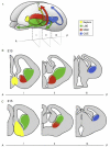
Figure 3.1
Figure 3.1
Schematic representation of the developing murine brain. (A) Three-dimensional view of a developing brain indicating the ventral anatomical regions that give rise to interneurons and their correspondent migratory pathways. The LGE (green) generates interneurons that migrate to the olfactory bulb and striatum. Both the MGE (red) and CGE (blue) produce cortical interneurons. Whether the septum produces cortical interneurons (yellow) is still a matter of debate. (B–C) Coronal views of the brain in (A) at three locations (i–iii) along the anterior (A)–posterior (P) axis at the embryonic ages E13 (B) and E15 (C).
Figure 3.2
Figure 3.2
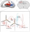
Differential origin of cortical interneuron subtypes. (A) Three-dimensional view of an embryonic murine brain highlighting the two ventral regions that produce cortical interneurons, the MGE (red) and the CGE (blue). (B) Coronal view of an adult brain, illustrating the proportion and relative distribution of cortical interneurons derived from the MGE (red dots) and the CGE (blue dots). (C) Detailed schematic view of the boxed region in (B) illustrating the main interneuron subtypes derived from the MGE (red cells) and CGE (blue cells), and how they characteristically interact with the pyramidal cells (black cells). This figure is adapted from Kawaguchi and Kubota (2002).
Figure 3.3
Figure 3.3
Temporal origin of cortical interneurons. (A) Examples of the morphological and electrophysiological diversity of cortical interneurons derived from the MGE at different times. Axons and dendrites are indicated in red and blue, respectively. This figure is adapted from Miyoshi et al. (2007). (B) Diagram of temporal origin of three subtypes of MGE interneurons, the somatostatin (SST—pink), the parvalbumin (PV—orange), and a subtype of CGE-derived interneuron (VIP—blue).
Figure 3.4
Figure 3.4
Cladistic hypothesis of cortical interneuron specification. We believe the fate of inhibitory neuron subtypes are sequentially influenced by both intrinsic genetic cues within the progenitor populations and environmental signals experienced by these populations postmitotically. In this model, we suggest that while the cardinal subdivisions are dependent on an intrinsic (genetic) program, the secondary subdivisions are determined later by environmental signals.
Figure 3.5
Figure 3.5
Summary of the essential events and processes that influence interneuron development. Embryonic ages are dominated by proliferation and cell migration. During this period GABA is excitatory. The first postnatal weeks are marked by early activity patterns, including synchronous plateau assemblies (SPAs) that are preceded by giant depolarizing potentials (GDPs). During this later period GABA switches from being excitatory to be inhibitory.

References

    1. Aboitiz F, Montiel J. Co-option of signaling mechanisms from neural induction to telencephalic patterning. Rev. Neurosci. 2007;18:311–342. - PubMed
    1. Alcamo EA, Chirivella L, Dautzenberg M, Dobreva G, Farinas I, Grosschedl R, McConnell SK. Satb2 regulates callosal projection neuron identity in the developing cerebral cortex. Neuron. 2008;57:364–377. - PubMed
    1. Allène C, Cattani A, Ackman JB, Bonifazi P, Aniksztejn L, Ben-Ari Y, Cossart R. Sequential generation of two distinct synapse-driven network patterns in developing neocortex. J. Neurosci. 2008;28:12851–12863. - PMC - PubMed
    1. Anderson SA, Eisenstat DD, Shi L, Rubenstein JL. Interneuron migration from basal forebrain to neocortex: Dependence on Dlx genes [see comment] Science. 1997a;278:474–476. - PubMed
    1. Anderson SA, Kaznowski CE, Horn C, Rubenstein JL, McConnell SK. Distinct origins of neocortical projection neurons and interneurons in vivo. Cereb. Corte. 2002;12:702–709. - PubMed

MeSH terms

LinkOut - more resources