. 2009 Jul 15;331(2):350-60.
doi: 10.1016/j.ydbio.2009.05.538. Epub 2009 May 8.

A conserved Six-Eya cassette acts downstream of Wnt signaling to direct non-myogenic versus myogenic fates in the C. elegans postembryonic mesoderm

Affiliations

A conserved Six-Eya cassette acts downstream of Wnt signaling to direct non-myogenic versus myogenic fates in the C. elegans postembryonic mesoderm

Nirav M Amin et al. Dev Biol. .

Abstract

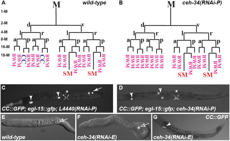
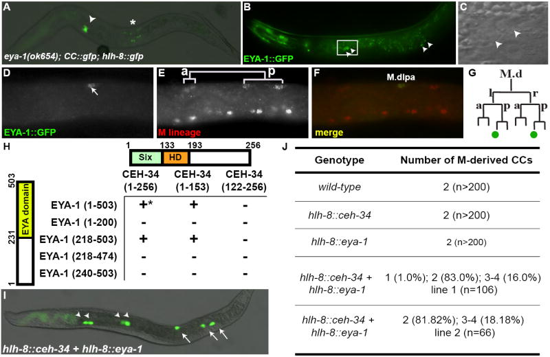
The subdivision of mesodermal cells into muscle and non-muscle cells is crucial to animal development. In the C. elegans postembryonic mesoderm, this subdivision is a result of an asymmetric cell division that leads to the formation of striated body wall muscles and non-muscle coelomocytes. Here we report that the Six homeodomain protein CEH-34 and its cofactor Eyes Absent, EYA-1, function synergistically to promote the non-muscle fate in cells also competent to form muscles. We further show that the asymmetric expression of ceh-34 and eya-1 is regulated by a combination of 1) mesodermal intrinsic factors MAB-5, HLH-1 and FOZI-1, 2) differential POP-1 (TCF/LEF) transcriptional activity along the anterior-posterior axis, and 3) coelomocyte competence factor(s). These factors are conserved in both vertebrates and invertebrates, suggesting a conserved paradigm for mesoderm development in metazoans.


Figures

Figure 1
Figure 1. ceh-34 is required for CC fates
All images in Figures 1-4 are ventral/lateral views with anterior to the left, unless otherwise noted. (A,B) Early M lineage in wild-type (A) and ceh-34(RNAi-P) (B) animals. Stages of M lineage (1-M to 18-M) are indicated. (C,D) L4440-RNAi treated control (C) and ceh-34(RNAi-P) (D) adults. CCs are visualized using intrinsic CC∷gfp, with embryonic CCs labeled with arrowheads and M-derived CCs with arrows. Type I vulval muscles are visualized using egl-15∷gfp, denoted by asterisks. M-derived CCs are missing in ceh-34(RNAi) animals (D). (E-F) L1 larva of water-injected (E) or ceh-34 dsRNA (F) injected animals. The M mesoblast is indicated by expression of hlh-8∷gfp (open arrow). (G) ceh-34(RNAi-E) adult with only one pair of embryonic CCs (arrowhead). M, M mesoblast; d, dorsal; v, ventral; l, left; r, right; a, anterior; p, posterior; CC, coelomocyte; BWM, body wall muscle; SM, sex myoblast.
Figure 2
Figure 2. ceh-34 is expressed in the M-derived CC precursors
(A) Representations of the ceh-34 translational reporter constructs: pNMA90 [ceh-34p∷ceh-34 genomic ORF∷gfp∷unc-54 3’UTR] and pNMA94 [ceh-34p∷gfp∷ceh-34 cDNA∷ceh-34 3’UTR]. The regions deleted in tm3330 and tm3733 are shown. (B-G): Representative images of live animals expressing ceh-34∷gfp (pNMA90) at different developmental stages. Two focal planes of a bean stage embryo (B,C, anterior to the left and dorsal up) and a 3-fold stage embryo (D,E). (F) A L1 larva and (G) an adult. Arrows in G denote BWMs. (H-O) The left side of a wild-type (H-K) or mab-5(RNAi-P) (L-O) L1 larva double labeled with CEH-34∷GFP (H,L) and anti-FOZI-1 antibody (I,M) at the 16-M stage. (J,N) Merged images of H and I, and L and M, respectively. CEH-34∷GFP was detected in M.dlpa (I) and M.drpa (not shown), both M-derived CC precursors. (K,O) Summary of ceh-34∷gfp expression in the M lineage, with ceh-34∷gfp-positive cells in green circles. All images are anterior to the left.
Figure 3
Figure 3. EYA-1 acts as a cofactor for CEH-34 in CC fate specification
(A) An eya-1(ok654) L4 larva missing one pair of embryonic CCs and both M-derived CCs. Arrowhead denotes the anterior pair of embryonically-derived CCs. GFP positive cells near the asterisk are SM descendants labeled by hlh-8∷gfp. (B,C) A wild-type adult expressing eya-1∷gfp (B). GFP was detected in the four embryonically-derived CCs (arrowheads). (C) Magnified DIC view of the anterior pair of CCs in the box in (B). (D-F) The left side of an L1 larva double labeled with EYA-1∷GFP (D), anti-FOZI-1 antibody (E) at the 16-M stage. (F) A merged image of D and E. EYA-1∷GFP was detected in M.dlpa (F) and M.drpa (not shown). (G) Summary of eya-1∷gfp expression in the M lineage, with eya-1∷gfp-positive cells in green circles. (H) The Six domain of CEH-34 binds to the Eya domain of EYA-1 via the yeast two-hybrid assay. Respective amino acid residues are labeled next to schematics of each protein. *All interactions were tested in both directions, with the exception of GBD-EYA-1(1-503), which auto-activates reporter expression. Western blot analysis was performed to ensure proper protein expression. (I,J) Forced expression of both ceh-34 and eya-1 in the M lineage leads to ectopic CC fates. Panel I shows an example of such a worm. Arrowheads point to embryonically-derived CCs while arrows point to M-derived CCs.
Figure 4
Figure 4. CEH-34 acts downstream of SYS-1 and POP-1 in the M lineage to specify CC fates
(A-H) Localization of POP-1∷GFP (A) and SYS-1∷GFP (E) in the M lineage of wild-type animals. (B,F) M lineage cells marked by anti-FOZI-1 staining; (C,G) the corresponding merged images. POP-1∷GFP is enriched in the nuclei of anterior cells (A), while SYS-1∷GFP is enriched in the nuclei of posterior cells (E). (D,H) Summary of asymmetric localization patterns of POP-1∷GFP and SYS-1∷GFP, respectively, in the M lineage. Black circles represent enriched localization; grey circles represent lower GFP levels. (I) A sys-1(q544) L4 larva with extra M-derived CCs (arrow) and its corresponding lineage (J). Arrowheads denote embryonically-derived CCs. (K-R) Two sys-1(RNAi) larvae with ectopic ceh-34∷gfp expression. K-M are lateral views while O-Q are dorsal images. (K,O) ceh-34∷gfp expression; (L,P) anti-FOZI-1 staining (with dorsal M lineage cells labeled); (M,Q) corresponding merged images; (N,R) corresponding lineages for K-M and O-Q, respectively, with green circles representing ceh-34∷gfp. (S-Z) POPTOP∷mCherry reporter expression in the M lineage of wild-type (S-V) and sys-1(RNAi-P) (W-Z) animals. (S,W) mCherry signal; (T,X) M lineage cells (hlh-8∷gfp); (U,Y) corresponding merged images; (V,Z) Summary of mCherry (red circles) expression in the M lineage. Note the faint mCherry signal in M.dlp and strong mCherry signal in M.vlpa and M.vlpp (U) in the wild-type animal, and the absence of mCherry signal in the M lineage of the sys-1(RNAi-P) animal (Y).
Figure 5
Figure 5. A model for non-muscle CC fate specification in the M lineage
(A) ceh-34 and eya-1 expression and subsequent specification of M-derived CC fate requires the combinatorial actions of three sets of factors: the M lineage intrinsic transcription factors including MAB-5, FOZI-1 and HLH-1 that are required for CC and BWM fate, the cell competence factor(s) X, and the repressive activity of POP-1 due to a low SYS-1 to POP-1 ratio. This low SYS-1 to POP-1 ratio leads to the repression of a repressor of ceh-34 and eya-1, which in turn results in the expression of ceh-34 and eya-1. (B) Combinatorial regulatory inputs leading to ceh-34 and eya-1 expression and CC fates. Solid lines in (A) and (B) do not represent direct regulation.

References

    1. Amin NM, Hu K, Pruyne D, Terzic D, Bretscher A, Liu J. A Zn-finger/FH2-domain containing protein, FOZI-1, acts redundantly with CeMyoD to specify striated body wall muscle fates in the Caenorhabditis elegans postembryonic mesoderm. Development. 2007;1:19–29. - PubMed
    1. Barolo S. Transgenic Wnt/TCF pathway reporters: all you need is Lef? Oncogene. 2006;57:7505–7511. - PubMed
    1. Brenner S. The genetics of Caenorhabditis elegans. Genetics. 1974;1:71–94. - PMC - PubMed
    1. Chen R, Amoui M, Zhang Z, Mardon G. Dachshund and eyes absent proteins form a complex and function synergistically to induce ectopic eye development in Drosophila. Cell. 1997;7:893–903. - PubMed
    1. Cheyette BN, Green PJ, Martin K, Garren H, Hartenstein V, Zipursky SL. The Drosophila sine oculis locus encodes a homeodomain-containing protein required for the development of the entire visual system. Neuron. 1994;5:977–996. - PubMed

Publication types

MeSH terms

LinkOut - more resources