Review
. 2009 May 9;373(9675):1632-44.
doi: 10.1016/S0140-6736(09)60371-8.

Intracerebral haemorrhage

Affiliations
Review

Intracerebral haemorrhage

Adnan I Qureshi et al. Lancet. .

Abstract

Intracerebral haemorrhage is an important public health problem leading to high rates of death and disability in adults. Although the number of hospital admissions for intracerebral haemorrhage has increased worldwide in the past 10 years, mortality has not fallen. Results of clinical trials and observational studies suggest that coordinated primary and specialty care is associated with lower mortality than is typical community practice. Development of treatment goals for critical care, and new sequences of care and specialty practice can improve outcome after intracerebral haemorrhage. Specific treatment approaches include early diagnosis and haemostasis, aggressive management of blood pressure, open surgical and minimally invasive surgical techniques to remove clot, techniques to remove intraventricular blood, and management of intracranial pressure. These approaches improve clinical management of patients with intracerebral haemorrhage and promise to reduce mortality and increase functional survival.


Figures

Figure 1
Figure 1. Cascade of neural injury initiated by intracerebral haemorrhage
The steps in the first 4 h are related to the direct effect of the haematoma, later steps to the products released from the haematoma. BBB=blood–brain barrier. MMP=matrix metallopeptidase. TNF=tumour necrosis factor. PMN=polymorphonuclear cells.
Figure 2
Figure 2. Progression of haemotoma and oedema on CT
Top: hyperacute expansion of haematoma in a patient with intracerebral haemorrhage on serial CT scans. Small haematoma detected in the basal ganglia and thalamus (A). Expansion of haematoma after 151 min (B). Continued progression of haematoma after another 82 min (C). Stabilisation of haematoma after another 76 min (D). Bottom: progression of haematoma and perihaematomal oedema in a patient with intracerebral haemorrhage on serial CT scans. The first scan (E) was acquired before the intracerebral haemorrhage. Perihaematoma oedema is highlighted in green to facilitate recognition of progression of oedema. At 4 h after symptom onset there is a small haematoma in the basal ganglia (F). Expansion of haematoma with extension into the lateral ventricle and new mass-effect and midline shift at 14 h (G). Worsening hydrocephalus and early perihaematomal oedema at 28 h (H). Continued mass-effect with prominent perihaematomal oedema at 73 h (I). Resolving haematoma with more prominent perihaematomal oedema at 7 days (J).
Figure 3
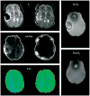
Figure 3. Advanced MRI of lobar intracerebral haemorrhage
Left: before craniotomy. Middle: after craniotomy for treatment of mass-effect and removal of haematoma. Sequential T2, lactate magnetic resonance spectroscopy, and perfusion studies showed qualitative decreases of perihaematomal oedema and perihaematomal lactate and increased occipital regional perfusion measured as time to peak of bolus injectate (TTP) after removal of clot; TTP is represented by intensity and distribution of green colour. Right: magnetic susceptibility images show paramagnetic influence before surgery and limited susceptibility after removal of the iron-containing blood clot by craniotomy. Figures provided by J Ricardo Carhuapoma (Johns Hopkins Medical Institution, Baltimore, MD, USA).
Figure 4
Figure 4. Detection of microhaemorrhages with MRI and CT scans
Left: asymptomatic pontine microbleed (arrow) in a patient with ischaemic stroke shown as focal hypointensity on gradient echo MRI. Right: microbleed not detected on CT scan. Figures are provided by David S Liebeskind (University of California at Los Angeles, CA, USA).
Figure 5
Figure 5
Management algorithm for patients with intracerebral haemorrhage
Figure 6
Figure 6. Odds ratio for death or disability in patients with lobar intracerebral haemorrhage treated surgically or conservatively
Boxes are Peto’s odds ratio (OR), lines are 95% CI. Adapted with permission from Lippincott Williams and Wilkins.

References

    1. Qureshi AI, Tuhrim S, Broderick JP, Batjer HH, Hondo H, Hanley DF. Spontaneous intracerebral hemorrhage. N Engl J Med. 2001;344:1450–60. - PubMed
    1. Labovitz DL, Halim A, Boden-Albala B, Hauser WA, Sacco RL. The incidence of deep and lobar intracerebral hemorrhage in whites, blacks, and hispanics. Neurology. 2005;65:518–22. - PubMed
    1. Sudlow CL, Warlow CP. Comparable studies of the incidence of stroke and its pathological types: results from an international collaboration. Stroke. 1997;28:491–99. - PubMed
    1. American Heart Organization. [accessed Nov 21, 2007];International cardiovascular disease statistics: cardiovascular disease (CVD) http://www.americanheart.org/downloadable/heart/1140811583642Internation....
    1. Qureshi AI, Suri MFK, Nasar A, et al. Changes in cost and outcome among US patients with stroke hospitalized in 1990 to 1991 and those hospitalized in 2000 to 2001. Stroke. 2007;38:2180–84. - PubMed

Publication types

MeSH terms