Comparative Study
. 2009 Jun 20;389(1-2):132-40.
doi: 10.1016/j.virol.2009.04.012. Epub 2009 May 9.

Vaccinia virus strain differences in cell attachment and entry

Affiliations
Comparative Study

Vaccinia virus strain differences in cell attachment and entry

Zain Bengali et al. Virology. .

Abstract

Vaccinia virus (VACV) strain WR can enter cells by a low pH endosomal pathway or direct fusion with the plasma membrane at neutral pH. Here, we compared attachment and entry of five VACV strains in six cell lines and discovered two major patterns. Only WR exhibited pH 5-enhanced rate of entry following neutral pH adsorption to cells, which correlated with sensitivity to bafilomycin A1, an inhibitor of endosomal acidification. Entry of IHD-J, Copenhagen and Elstree strains were neither accelerated by pH 5 treatment nor prevented by bafilomycin A1. Entry of the Wyeth strain, although not augmented by pH 5, was inhibited by bafilomycin A1. WR and Wyeth were both relatively resistant to the negative effects of heparin on entry, whereas the other strains were extremely sensitive due to inhibition of cell binding. The relative sensitivities of individual vaccinia virus strains to heparin correlated inversely with their abilities to bind to and enter glycosaminoglycan-deficient sog9 cells but not other cell lines tested. These results suggested that that IHD-J, Copenhagen and Elstree have a more limited ability than WR and Wyeth to use the low pH endosomal pathway and are more dependent on binding to glycosaminoglycans for cell attachment.


Figures

Fig. 1
Fig. 1
Entry of of VACV strains. (A) BS-C-1 cells were incubated with VACV recombinant luc strains at 4°C at neutral pH for 1 h at a multiplicity of 1 plaque forming unit per cell, followed by washing to remove unbound virus and exposure to pH 5 (black bars) or pH 7.4 (gray bars) buffer for 3 min at 37°C. Cells were then washed and incubated at 37°C at neutral pH for 1 h. Cells were lysed and luc activity measured. (B) Fold enhancement of low pH was calculated by dividing the low pH relative luminescent units (RLU) by the neutral pH RLU in panel A. Error bars are plotted in each case, though in some the differences are too small to be resolved.
Fig. 2
Fig. 2
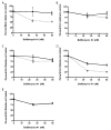
Inhibition of VACV entry with endosomal acidification inhibitor bafilomycin A1. BS-C-1 cells were pretreated with bafilomycin A1 for 1 h at 37°C. Pretreated cells were then incubated with (A) WR, (B) IHD-J, (C) Copenhagen, (D) Wyeth, and (E) Elstree luc recombinant strains of VACV in the presence of bafilomycin A1 at neutral pH for 1 h at 4°C. Unattached virus was removed, and the cells were washed and then exposed to pH 5 (filled squares, solid line) or pH 7.4 (open squares, dashed line) buffer for 3 min at 37°C. Cells were then washed and incubated at 37°C at neutral pH for 1 h in the presence of bafilomycin A1. Untreated cells exposed to virus and the respective buffers were utilized as controls. Error bars are plotted for each point, though in some the differences are too small to be resolved.
Fig. 3
Fig. 3
Effect of low pH treatment prior to adsorption on entry of VACV strains. (A) VACV strains were pre-treated with pH 5 (black bars) or pH 7.4 (gray bars) for 3 min. Treated virus was incubated on BS-C-1 cells for 1 h at 4°C at neutral pH. After washing, cells were incubated at 37°C for 1 h and luc values were measured. (B) The fold enhancement of luc activity at low pH over neutral pH was calculated from data in panel A. Error bars are plotted in each case, though in some the differences are too small to be resolved.
Fig. 4
Fig. 4
Entry of VACV in different cell types. The indicated cell types were incubated with WR (A, C) and IHD-J (B, D) at neutral pH at 4°C. The cells were then washed and treated with pH 5 (black bars) or pH 7.4 (gray bars) buffer for 3 min at 37°C. The pH was neutralized and the cells were incubated in regular medium for 1 h and luc was measured (A, B). The fold enhancement of low pH treatment was calculated (C, D). Error bars are plotted in each case, though in some the differences are too small to be resolved.
Fig. 5
Fig. 5
Effects of soluble GAGs on VACV entry. WR (A, C, E) and IHD-J (B, D, F) strains of VACV were pretreated with 50 μg per ml of heparin (HP), heparan sulfate (HS), chondroitin sulfate (CS) and combinations of HP+HS and HP+CS for 30 min on ice. Without removal of the soluble GAGs, the treated virus was then incubated with BS-C-1 cells at 4°C for 1 h at neutral pH followed by a wash and treatment with (A, B) pH 7.4 buffer or (C, D) pH 5 buffer. Cells were then washed and incubated for 1 h at 37°C at neutral pH, lysed and luc measured. (E, F) Percent of control (virus mock incubated with GAGs) for each treatment was also determined from the data in panels A–D. Error bars are plotted in each case, though in some the differences are too small to be resolved.
Fig. 6
Fig. 6
Effect of heparin concentration on VACV entry. WR (A) and IHD-J (B) were incubated with 0 to 50 μg/ml of heparin for 30 min on ice and added to BS-C-1 cells at 4°C for 1 h. Cells were then washed and incubated with pH 5 or pH 7.4 buffer for 3 min at 37°C followed by washing and incubation in regular medium at 37°C for 1 h. Cells were then lysed and luc activity measured. Error bars are plotted for each point, though in some the differences are too small to be resolved.
Fig. 7
Fig. 7
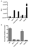
Effect of heparin on entry of VACV strains. (A) VACV strains were treated with 50 μg/ml of heparin (white bars) or mock treated (black bars) for 30 min on ice. Treated virus was then added to cells and incubated at 4°C for 1 h. Cells were then washed and incubated at 37°C for 1 h followed by lysis and measurement of luc. (B) The percent RLU relative to control of treated virus was also calculated. Error bars are plotted in each case, though in some the differences are too small to be resolved.
Fig 8
Fig 8
Effect of laminin on entry of VACV. WR (A, C) and IHD-J (B, D) strains of VACV were pretreated with 50 μg per ml of laminin (LN), HP and a combination of HP+LN. (A, B) Treated virus was then incubated with BS-C-1 cells at 4°C for 1 h followed by a wash and treatment with pH 7.4 (gray bars) buffer or pH 5 buffer (black bars). Cells were then washed and incubated for 1 h at 37°C, lysed and luc measured. (C, D) Percent of control (virus mock incubated) for each treatment was also determined from the data in panels A, B. Error bars are plotted in each case, though in some the differences are too small to be resolved.
Fig. 9
Fig. 9
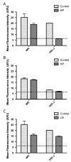
Effect of heparin and laminin on binding of WR and IHD-J to cells. Recombinant WR and IHD-J with GFP fused to the VACV A4 core protein were incubated with or without heparin (A, B) or laminin (C) and adsorbed to HeLa (A, C) or sog9 (B) cells at 4°C for 1 h. Unbound virus was removed by washing and the cells were harvested by scraping. The amounts of virus bound to the cells were determined by flow cytometry and the ratios of mean fluorescence determined. The dashed lines indicate background fluorescence. Error bars are plotted in each case, though in some the differences are too small to be resolved.

References

    1. Armstrong JA, Metz DH, Young MR. The mode of entry of vaccinia virus into L cells. J Gen Virol. 1973;21:533–537. - PubMed
    1. Banfield BW, Leduc Y, Esford L, Schubert K, Tufaro F. Sequential isolation of proteoglycan synthesis mutants by using herpes simplex virus as a selective agent: evidence for a proteoglycan-independent virus entry pathway. J Virol. 1995;69:3290–3298. - PMC - PubMed
    1. Bisht H, Weisberg AS, Moss B. Vaccinia virus l1 protein is required for cell entry and membrane fusion. J Virol. 2008;82:8687–8694. - PMC - PubMed
    1. Blasco R, Sisler JR, Moss B. Dissociation of progeny vaccinia virus from the cell membrane is regulated by a viral envelope glycoprotein: effect of a point mutation in the lectin homology domain of the A34R gene. J Virol. 1993;67:3319–3325. - PMC - PubMed
    1. Brown E, Senkevich TG, Moss B. Vaccinia virus F9 virion membrane protein is required for entry but not virus assembly, in contrast to the related L1 protein. J Virol. 2006;80:9455–9464. - PMC - PubMed

Publication types