Review
. 2009 Jun;30(6):269-77.
doi: 10.1016/j.tips.2009.03.002. Epub 2009 May 8.

Emerging antiviral targets for influenza A virus

Affiliations
Review

Emerging antiviral targets for influenza A virus

Robert M Krug et al. Trends Pharmacol Sci. 2009 Jun.

Abstract

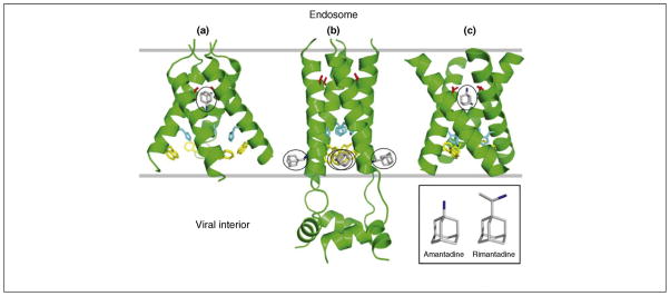
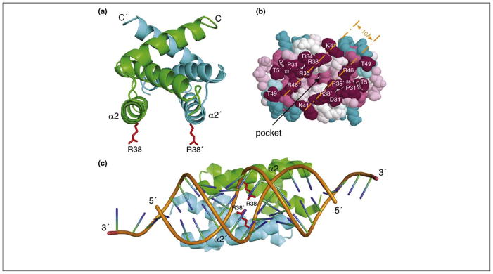
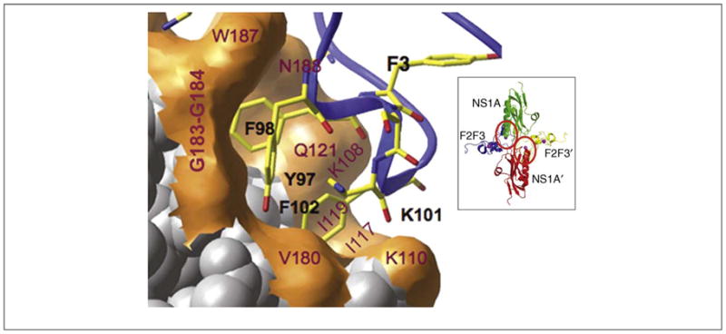
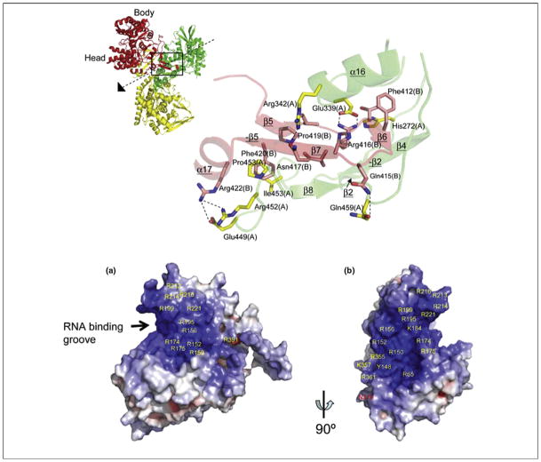
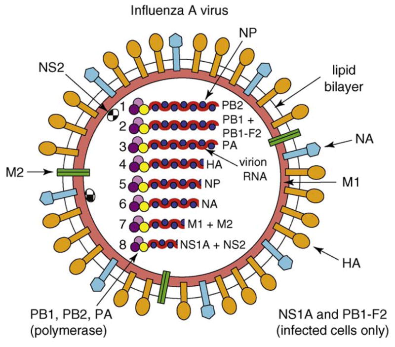
The potential threat of a pandemic caused by H5N1 influenza A viruses has stimulated increased research on developing new antivirals against influenza A viruses. Current antivirals are directed against the M2 protein (named adamantanes) and the neuraminidase (named zanamivir and oseltamivir). However, both seasonal and H5N1 influenza A viruses have developed resistance to adamantanes and oseltamivir. Accordingly, new antivirals directed at the M2 and neuraminidase proteins, and against the hemagglutinin protein, are being developed. In addition, elucidation of the structural basis for several crucial functions of other viral proteins (specifically the non-structural NS1A protein, the nucleoprotein and the viral polymerase) has identified novel targets for the development of new antivirals. Here, we describe how functional and structural studies led to the discovery of these novel targets and also how structural information is facilitating the rational design of new drugs against previously identified targets.


Figures

Figure 1
Figure 1
Current structures of the influenza A virus M2 protein channel in complex with either amantadine or rimantadine. (a) X-ray structure (3.5 Å resolution) of the TM domain of M2 (residues 22–46) tetramer in complex (1:1) with amantadine in octyl-β-D-glucopyranoside detergent, pH 5.3 [27]. (b) Solution NMR structure of the TM domain and additional C-terminal region of M2 (residues 18–60) tetramer in complex (1:4) with rimantadine in dihexanoyl phosphatidylcholine detergent micelles, pH 7.5 [28]. The four rimantadine molecules are outside the channel. (c) Solid-state NMR structure of the TM domain of M2 (residues 22–46) tetramer in complex (1:1) with amantadine in dilauroyl phosphatidylcholine bilayers, pH 7.5 [29]. For each structure, side chains of Ser31 (red), His37 (cyan) and Trp41 (yellow) are shown, along with bound amanatadine or rimantadine (grey with nitrogen in blue; circled).
Figure 2
Figure 2
Potential antiviral target in the NS1A dsRNA-binding domain. (a) Ribbon representation of the solution NMR structure of Ud NS1A(1–73) [53]. NS1A and NS1A′ subunits are shown in green and cyan, respectively, and the side chains of Arg38 and Arg38′ (R38 and R38′) are in red. α2 and α2′ denote the antiparallel helices; C stands for C terminus. (b) Space-filling model of the Ud NS1A(1–73), showing tracks of conserved residues on the RNA-binding face of the protein [55]. Residues are colored according to their degree of conservation across the entire NS1A family, ranging from highest (purple) to lowest (cyan). The separation across the basic edges of the conserved tracks corresponds to 10 Å, the width of the major groove in dsRNA. The pocket on the RNA-binding face is indicated. Reproduced, with permission, from Ref. [55]. (c) The crystal structure of NS1A(1–70) from influenza A/Puerto Rico/8/34 (H1N1) in complex with a 19-bp dsRNA duplex [56]. Ribbon representation of NS1A(1–70)–dsRNA showing the major grove mode of binding in the complex. NS1A RBD subunits are colored as in panel (a); the dsRNA backbone is orange. Reproduced, with permission, from Ref. [56].
Figure 3
Figure 3
Potential antiviral target in the NS1A effector domain. Expanded view of the F2F3-binding pocket of the Ud NS1A ED [61]. NS1A residues labeled red interact with aromatic side chains in the F3 zinc finger. Inset: ribbon representation of the Ud NS1A(85–215)–F2F3 complex, comprising two NS1A effector domains (green and red) and two F2F3 zinc finger domains (blue and yellow). The F2F3-binding pockets in the two NS1A subunits of the tetramer are circled. Modified, with permission, from Ref. [61].
Figure 4
Figure 4
Potential antiviral targets in NP. Crystal structure of the NP from HINI influenza A/WSN/33 [66]. (a) Interactions between subunit A (green) and the tail loop of subunit B (red) from the trimeric structure of NP shown on the left. For each denoted amino acid, the subunit in which it is located (A or B) is shown in a parenthesis. (b) Electrostatic surface potential (blue, positive; red, negative) of subunit B from NP, identifying a putative RNA-binding site. Reproduced, with permission, from Ref. [66].
Figure 5
Figure 5
Potential antiviral target in the viral polymerase. Structure of the PAC–PB1N complex. Surface image showing the hydrophobic contacts between PA (green) and the N terminus of PB1 (orange) in the crystal structure of PA(239–716)–PB1(1–81) from influenza A/Puerto Rico/8/1934 (H1N1) [68]. All other residues in PA are colored blue. Reproduced, with permission, from Ref. [68].
Figure I
Figure I
Schematic representation of influenza A virus.

References

    1. Lamb RA, Krug RM. Orthomyxoviridae: the viruses and their replication. In: Knipe DM, Howley PM, editors. Fields Virology. 4. Lippincott Williams & Wilkins; 2001. pp. 1487–1532.
    1. Wright PF, Webster RG. Orthomyxoviruses. In: Knipe DM, Howley PM, editors. Fields Virology. 4. Lippincott Williams & Wilkins; 2001. pp. 1533–1579.
    1. Reid AH, et al. The 1918 Spanish influenza: integrating history and biology. Microbes Infect. 2001;3:81–87. - PubMed
    1. Horimoto T, Kawaoka Y. Influenza: lessons from past pandemics, warnings from current incidents. Nat Rev Microbiol. 2005;3:591–600. - PubMed
    1. Noah DL, Krug RM. Influenza virus virulence and its molecular determinants. In: Maramorsch K, Shatkin AJ, editors. Advances in Virus Research. Vol. 65. Elsevier; 2005. pp. 121–145. - PubMed

Publication types

MeSH terms