Review
. 2009 Jun;33(6):843-63.
doi: 10.1016/j.neubiorev.2009.02.004. Epub 2009 Feb 24.

Neural bases of eye and gaze processing: the core of social cognition

Affiliations
Review

Neural bases of eye and gaze processing: the core of social cognition

Roxane J Itier et al. Neurosci Biobehav Rev. 2009 Jun.

Abstract

Eyes and gaze are very important stimuli for human social interactions. Recent studies suggest that impairments in recognizing face identity, facial emotions or in inferring attention and intentions of others could be linked to difficulties in extracting the relevant information from the eye region including gaze direction. In this review, we address the central role of eyes and gaze in social cognition. We start with behavioral data demonstrating the importance of the eye region and the impact of gaze on the most significant aspects of face processing. We review neuropsychological cases and data from various imaging techniques such as fMRI/PET and ERP/MEG, in an attempt to best describe the spatio-temporal networks underlying these processes. The existence of a neuronal eye detector mechanism is discussed as well as the links between eye gaze and social cognition impairments in autism. We suggest impairments in processing eyes and gaze may represent a core deficiency in several other brain pathologies and may be central to abnormal social cognition.


Figures
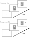
Fig. 1
Fig. 1
Typical orienting-to-gaze paradigm. A central face cue with averted gaze is presented prior to target onset. Although the cue does not predict the location of the target, subjects respond faster to targets when gaze direction and target location match (congruent trials) and slower when they do not match (incongruent trials).
Fig. 2
Fig. 2
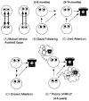
(A–E) Schematic descriptions of the various social situations involving the use of gaze direction. The approximate ages at which the various capabilities emerge are in parenthesis. Adapted from Emery (2000), with permission.
Fig. 3
Fig. 3
The ERP component N170 recorded at a right cerebellar electrode (CB2) for faces and isolated eyes (adapted from Itier et al., 2006b). The N170 is larger and delayed for eyes compared to faces. The respective topographies, representing the voltage distribution on the scalp at the peak of the N170 for each category, are also different, reflecting different underlying generators.
Fig. 4
Fig. 4
The N170 and M170 components obtained after presentation of a face. Topographies are shown at the peak of the components. In one study (Itier et al., 2006a), the M170 recorded with MEG generated a right fusiform gyrus (FG) source and a bilateral source within the inferior and medial occipital gyri (IOG/MOG) when analyzed with the beam former technique event-related Synthetic Aperture Magnetometry (er-SAM). Using the 3D current density method LAURA, Batty and Taylor (2003) and Itier and Taylor (2004a) found that the N170 recorded with ERPs was best modeled by a bilateral source within the STS region. In contrast, in most dipole source analysis such as the one performed by Itier and Taylor (2002) using brain evoked source analysis (BESA), the N170 is often best modeled by dipoles within the FG. These findings led to the hypothesis that approximately 170 ms after a face onset, three different sources are active: the FG, the STS and the IOG/MOG. However, in some cases that remain to be determined, the STS would be best recorded with ERPs due to the proximity of the source to the scalp (underneath temporo-parietal sites) and to a possible radial orientation. The FG and IOG/MOG sources would be best captured with MEG if the sources are tangential. However the sources in the FG may be composed of both tangential and radial components, explaining why sometimes the N170 is modeled by sources in the FG.
Fig. 5
Fig. 5
(A) Intracranial data in humans showed that patches of cortex within the fusiform gyrus (FG, axial view) and superior temporal sulcus (STS, lateral view) are selective for whole faces and for facial components such as eyes (Courtesy of Dr. A. Puce). (B) Effects of inversion on the N170 (shown here at a right cerebellar electrode—CB2) in an orientation discrimination task for isolated eyes, faces, face-without-eyes and houses from Itier et al. (2007b). Horizontal lines show that the N170 amplitude for faces-without-eyes, presented upright or inverted, did not differ from that to upright normal faces. In contrast, inverting full faces increased the N170, which amplitude was not different from that recorded to upright or inverted eyes. This suggests that inverted faces are processed like isolated eyes and that the inversion effect on the N170 is driven by the presence of the eyes. (C) Simplified neural model of early face processing adapted from Itier et al. (2007b). It is assumed that the N170 arises from the FG and the STS region, in which face-selective and eye-selective neurons likely co-exist. The ‘+’ signs signify the neurons are active. Both face- and eye-selective neurons respond to isolated eyes. However, only face-selective neurons respond to faces. The ‘−’ sign followed by a question mark indicates a possible inhibition mechanism from the face neurons onto the eye neurons which would thus not respond to the eyes within an upright face configuration. Regardless of whether they are simply not activated or inhibited by the face neurons, the eye-selective neurons do not respond to the eyes of the face because of the facial context (configuration). When the face is inverted, the facial configuration is disrupted and the eye-selective neurons now respond, just like for isolated eyes, producing the N170 amplitude increase. Note that “face-selective” neurons are sensitive to the face configuration: although they respond to inverted faces and faces-without-eyes, their response is delayed compared to normal full upright faces (see text and Itier et al., 2007b for a complete description).

References

    1. Abell F, Krams M, Ashburner J, Passingham R, Friston K, Frackowiak R, et al. The neuroanatomy of autism: a voxel-based whole brain analysis of structural scans. Neuroreport. 1999;10 (8):1647–1651. - PubMed
    1. Adams RB, Jr, Gordon HL, Baird AA, Ambady N, Kleck RE. Effects of gaze on amygdala sensitivity to anger and fear faces. Science. 2003;300 (5625):1536. - PubMed
    1. Adams RB, Jr, Kleck RE. Perceived gaze direction and the processing of facial displays of emotion. Psychol Sci. 2003;14 (6):644–647. - PubMed
    1. Adams RB, Jr, Kleck RE. Effects of direct and averted gaze on the perception of facially communicated emotion. Emotion. 2005;5 (1):3–11. - PubMed
    1. Adolphs R. Social cognition and the human brain. Trends Cogn Sci. 1999;3 (12):469–479. - PubMed

Publication types