

Review
doi: 10.1016/j.bbr.2009.03.004.
Epub 2009 Mar 17.
The functional neuroanatomy of depression: distinct roles for ventromedial and dorsolateral prefrontal cortex
Affiliations
- PMID: 19428640
- PMCID: PMC2680780
- DOI: 10.1016/j.bbr.2009.03.004
Item in Clipboard
Review
The functional neuroanatomy of depression: distinct roles for ventromedial and dorsolateral prefrontal cortex
Behav Brain Res.
.
Abstract
A primary aim in the neuroscientific study of depression is to identify the brain areas involved in the pathogenesis of symptoms. In this review, we describe evidence from studies employing various experimental approaches in humans (functional imaging, lesion method, and brain stimulation) that converge to implicate the ventromedial and dorsolateral sectors of prefrontal cortex as critical neural substrates for depression, albeit with distinct functional contributions. The putative roles of ventromedial and dorsolateral prefrontal cortex in depression are discussed in light of the results.
Figures
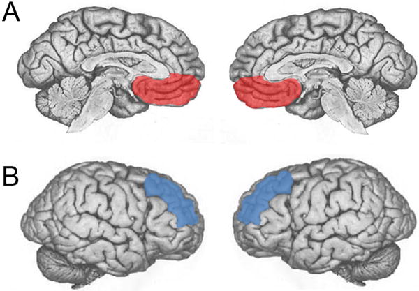
A. Depiction of vmPFC (in red) in midline views of each hemisphere. B. Depiction of dlPFC (in blue) in lateral views of each hemisphere.
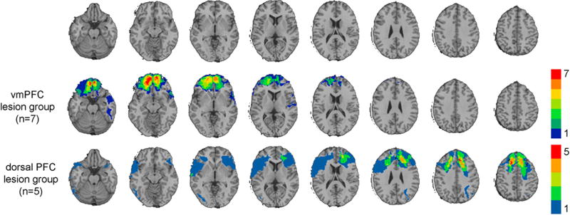
Lesion overlaps of VHIS patients with bilateral vmPFC or dlPFC damage. Color indicates the number of overlapping lesions at each voxel. Top row: Axial slices of a normal healthy brain, for reference. Second row: Lesion overlap for the vmPFC lesion group. Third row: Lesion overlap for the dorsal PFC lesion group.
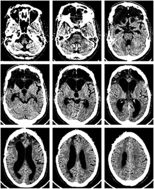
CT image (axial slices) of patient experiencing depression alleviation following suicide attempt. Ventral PFC is almost entirely destroyed, including vmPFC bilaterally (top row), whereas dorsal PFC is largely intact (bottom row).
References
-
- Davidson RJ, et al. Depression: perspectives from affective neuroscience. Annu Rev Psychol. 2002;53:545–74. - PubMed
-
- Drevets WC. Functional neuroimaging studies of depression: the anatomy of melancholia. Annu Rev Med. 1998;49:341–61. - PubMed
-
- Drevets WC. Orbitofrontal cortex function and structure in depression. Ann N Y Acad Sci. 2007;1121:499–527. - PubMed
-
- Kringelbach ML, Rolls ET. The functional neuroanatomy of the human orbitofrontal cortex: evidence from neuroimaging and neuropsychology. Prog Neurobiol. 2004;72(5):341–72. - PubMed
-
- Rosenkilde CE. Functional heterogeneity of the prefrontal cortex in the monkey: a review. Behav Neural Biol. 1979;25(3):301–45. - PubMed
Publication types
MeSH terms
Grants and funding
LinkOut - more resources
Full Text Sources
Other Literature Sources
Medical
