

Detection and quantification of botulinum neurotoxin type a by a novel rapid in vitro fluorimetric assay
- PMID: 19429547
- PMCID: PMC2704824
- DOI: 10.1128/AEM.00091-09
Detection and quantification of botulinum neurotoxin type a by a novel rapid in vitro fluorimetric assay
Abstract
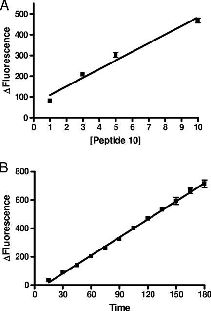
Botulinum neurotoxin type A (BoNT/A), the most poisonous substance known to humans, is a potential bioterrorism agent. The light-chain protein induces a flaccid paralysis through cleavage of the 25-kDa synaptosome-associated protein (SNAP-25), involved in acetylcholine release at the neuromuscular junction. BoNT/A is widely used as a therapeutic agent and to reduce wrinkles. The toxin is used at very low doses, which have to be accurately quantified. With this aim, internally quenched fluorescent substrates containing the fluorophore/repressor pair pyrenylalanine (Pya)/4-nitrophenylalanine (Nop) were developed. Nop and Pya were, respectively, introduced at positions 197 and 200 of the cleavable fragment (amino acids 187 to 203) of SNAP-25 (with norleucine at position 202 [Nle(202)]), which is acetylated at its N terminus and amidated at its C terminus. Cleavage of this peptide occurred between positions 197 and 198, as in SNAP-25, and was easily quantified by the strong fluorescence emission of the metabolite. To increase the assay sensitivity, the peptide sequence of the previous substrate was lengthened to account for exosite binding to BoNT/A. We synthesized the peptide PL50 (SNAP-25-NH(2) acetylated at positions 156 to 203 [Nop(197), Pya(200), Nle(202)]) and its analogue PL51, in which all methionines were replaced by nonoxidizable Nle. Consistent with a large increase in affinity for BoNT/A, PL50 and PL51 exhibit catalytic efficiencies of 2.6 x 10(6) M(-1) s(-1) and 8.85 x 10(6) M(-1) s(-1), respectively, and behave as the best fluorigenic substrates of BoNT/A reported to date. Under optimized assay conditions, they allow simple quantification of as little as 100 and 60 pg of BoNT/A, respectively, within 2 h with a classical fluorimeter. Calibration of the method against the mouse 50% lethal dose assay unequivocally validates the enzymatic assay.
Figures

Similar articles
-
Comparison of fluorigenic peptide substrates PL50, SNAPTide, and BoTest A/E for BoNT/A detection and quantification: exosite binding confers high-assay sensitivity.J Biomol Screen. 2013 Jul;18(6):726-35. doi: 10.1177/1087057113476089. Epub 2013 Feb 20. J Biomol Screen. 2013. PMID: 23427044
-
A substrate sensor chip to assay the enzymatic activity of Botulinum neurotoxin A.Biosens Bioelectron. 2013 Nov 15;49:276-81. doi: 10.1016/j.bios.2013.05.032. Epub 2013 May 29. Biosens Bioelectron. 2013. PMID: 23787358
-
High-throughput fluorogenic assay for determination of botulinum type B neurotoxin protease activity.Anal Biochem. 2001 Apr 15;291(2):253-61. doi: 10.1006/abio.2001.5028. Anal Biochem. 2001. PMID: 11401299
-
8-Hydroxyquinoline and hydroxamic acid inhibitors of botulinum neurotoxin BoNT/A.Curr Top Med Chem. 2014;14(18):2094-102. doi: 10.2174/1568026614666141022095114. Curr Top Med Chem. 2014. PMID: 25335884 Review.
-
Mass Spectrometric Detection of Bacterial Protein Toxins and Their Enzymatic Activity.Toxins (Basel). 2015 Aug 31;7(9):3497-511. doi: 10.3390/toxins7093497. Toxins (Basel). 2015. PMID: 26404376 Free PMC article. Review.
Cited by
-
Preferential entry of botulinum neurotoxin A Hc domain through intestinal crypt cells and targeting to cholinergic neurons of the mouse intestine.PLoS Pathog. 2012;8(3):e1002583. doi: 10.1371/journal.ppat.1002583. Epub 2012 Mar 15. PLoS Pathog. 2012. PMID: 22438808 Free PMC article.
-
Optimization of SNAP-25 and VAMP-2 Cleavage by Botulinum Neurotoxin Serotypes A-F Employing Taguchi Design-of-Experiments.Toxins (Basel). 2019 Oct 11;11(10):588. doi: 10.3390/toxins11100588. Toxins (Basel). 2019. PMID: 31614566 Free PMC article.
-
Proteomic Methods of Detection and Quantification of Protein Toxins.Toxins (Basel). 2018 Feb 28;10(3):99. doi: 10.3390/toxins10030099. Toxins (Basel). 2018. PMID: 29495560 Free PMC article. Review.
-
Substrates and controls for the quantitative detection of active botulinum neurotoxin in protease-containing samples.Anal Chem. 2013 Jun 4;85(11):5569-76. doi: 10.1021/ac4008418. Epub 2013 May 22. Anal Chem. 2013. PMID: 23656526 Free PMC article.
-
Sensing the deadliest toxin: technologies for botulinum neurotoxin detection.Toxins (Basel). 2010 Jan;2(1):24-53. doi: 10.3390/toxins2020024. Epub 2010 Jan 7. Toxins (Basel). 2010. PMID: 22069545 Free PMC article.
References
-
- Anne, C., F. Cornille, C. Lenoir, and B. P. Roques. 2001. High-throughput fluorigenic assay for determination of botulinum type B neurotoxin protease activity. Anal. Biochem. 291:253-261. - PubMed
-
- Arnon, S. S., R. Schechter, T. V. Inglesby, D. A. Henderson, J. G. Bartlett, M. S. Ascher, E. Eitzen, A. D. Fine, J. Hauer, M. Layton, S. Lillibridge, M. T. Osterholm, T. O'Toole, G. Parker, T. M. Perl, P. K. Russell, D. L. Swerdlow, and K. Tonat. 2001. Botulinum toxin as a biological weapon: medical and public health management. JAMA 285:1059-1070. - PubMed
-
- Blitzer, A., M. F. Brin, M. S. Keen, and J. E. Aviv. 1993. Botulinum toxin for the treatment of hyperfunctional lines of the face. Arch. Otolaryngol. Head Neck Surg. 119:1018-1022. - PubMed
-
- Breidenbach, M. A., and A. T. Brunger. 2004. Substrate recognition strategy for botulinum neurotoxin serotype A. Nature 432:925-929. - PubMed
Publication types
MeSH terms
Substances
LinkOut - more resources
Full Text Sources
Other Literature Sources
Medical
