Case Reports
. 2009 Sep;93(9):1234-40.
doi: 10.1136/bjo.2008.153577. Epub 2009 May 7.

Cellular origin of fundus autofluorescence in patients and mice with a defective NR2E3 gene

Affiliations
Case Reports

Cellular origin of fundus autofluorescence in patients and mice with a defective NR2E3 gene

N-K Wang et al. Br J Ophthalmol. 2009 Sep.

Abstract

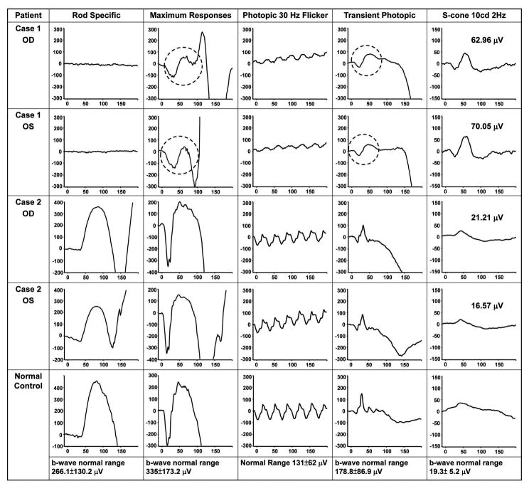
Aim: To characterise new clinical features in a family with enhanced S-cone syndrome (ESCS) and investigate the pathogenesis of these clinical features in the homozygous Nr2e3(rd7) (rd7) mutant mice.

Methods: Four patients from an affected family were included for genotypic and phenotypic study. Eye tissues from rd7 mice were used to detect a possible relationship between macrophages and autofluorescent material by immunohistochemistry (IHC) staining.

Results: Homozygous mutation in R311Q in NR2E3 was detected in this family. Colour photographs revealed that white dots do not correlate to hyperautofluorescent spots seen in autofluorescence imaging of the macula. OCT showed rosette-like lesions similar to those found in rd7 mice histology sections. From IHC analysis, we observed that F4/80 (a pan macrophage marker) and autofluorescence were colocalised to the same cells within the retina rosettes.

Conclusions: The retinal structure of a young ESCS patient with homozygous R311Q mutation in the NR2E3 gene is similar to that seen in the rd7 mice. The macrophages were found to contain autofluorescent materials in the retinal rosettes of rd7 mice. These data are consistent with macrophage infiltration contributing to the hyperautofluorescent spots found in our patients.


Figures

Figure 1
Figure 1
(A). Pedigree of a family in which mutation in the NR2E3 gene were identified. A mutant allele is indicated by (−); a normal allele is indicated by (+). Arrow shows the proband (1, homozygous), and the other members are heterozygous. (B). Direct DNA sequencing from proband’s consanguineous parents and brother showed heterozygous base-substitution at 932G→A in exon 6. (C). Direct sequencing from the proband revealed a homozygous base-substitution at 932G→A with a predicted missense mutation of R311Q (CGG to CAG).
Figure 2
Figure 2
(A). Composite fundus photographs of the right and left eyes of proband (case 1) showing multiple subretinal white deposits and prominent lesions in the macula (red arrows). (B). Composite fundus imaging of the right and left eyes of case 2 exhibited a normal retina without subretinal deposit. There are some artifacts arising from photography (yellow arrows illustrate examples of artifacts from the camera lens, blue arrows show reflections of the nerve fiber layer).
Figure 3
Figure 3
(A). Fundus autofluorescence exam (FAF) of proband (case 1) showed hyper-autofluorescent spots in the macular area and in the mid-peripheral retina. (B). Optical coherence tomography (OCT) 1, 2, 3, and 4 correspond to scanning sections in fig A. 1 showed cystic changes around the foveal lesion (OD). Note that the rosette-like lesions (arrow head) correspond to hyper-autofluorescent spots and loss of retinal lamination (2, 3 and 4). (C). FAF of case 2 showed several hyper-autofluorescent spots around the disc and arcades. (D). (5,6) OCT sectioning of case 2 (line 5 and 6 from fig C) showed normal retinal lamination without rosette-like lesions. Some shadows casted by retinal vessels are observed.
Figure 4
Figure 4
Electroretinogram (ERG) from case 1 revealed three characteristic findings: 1). no rod responses; 2) the waveforms of scotopic maximal response are very identical to the transient photopic responses except the size (circles); 3) the amplitude of a wave in the transient photopic response is larger than amplitude of photopic 30 Hz flicker. S-cone-specific ERG was of remarkably high amplitude (60–70 μV) and further confirmed the diagnosis of ESCS. ERG from case 2 was normal.
Figure 5
Figure 5
Color fundus photographs from wild type mouse (A) showed normal retina, whereas image from rd7 mouse (B) showed numerous subretinal white dots. Whole mount eyecup from wild type mouse (C) did not reveal hyperautofluorescence spots whereas image from rd7 mouse (D) showed hyperautofluorescence spots. (E). Spectral fluorescence of hyperautofluorescence spots from rd7 mouse retina demonstrated the wave length of highest fluorescence intensity was 540 nm to 560 nm when excited with light of 436 nm.
Figure 6
Figure 6
(A). Immunohistochemistry of a frozen section from rd7 mouse retina using anti-F4/80 antibodies (macrophage marker) showed one macrophage in the retinal rosette, or whorl lesion. (B). High magnification of white dots lined area from (A) showed images merge from different fluorescence channels. The autofluorescence (red) was co-localized with macrophage marker (green). INL: inner nuclear layer; ONL: outer nuclear layer; AF: autofluorescence; F4/80: macrophage marker; DAP: DAPI staining.

References

    1. Chen J, Rattner A, Nathans J. The rod photoreceptor-specific nuclear receptor Nr2e3 represses transcription of multiple cone-specific genes. J Neurosci. 2005;25:118–29. - PMC - PubMed
    1. Sharon D, Sandberg MA, Caruso RC, Berson EL, Dryja TP. Shared mutations in NR2E3 in enhanced S-cone syndrome, Goldmann-Favre syndrome, and many cases of clumped pigmentary retinal degeneration. Arch Ophthalmol. 2003;121:1316–23. - PubMed
    1. Haider NB, Jacobson SG, Cideciyan AV, et al. Mutation of a nuclear receptor gene, NR2E3, causes enhanced S cone syndrome, a disorder of retinal cell fate. Nat Genet. 2000;24:127–31. - PubMed
    1. Milam AH, Rose L, Cideciyan AV, et al. The nuclear receptor NR2E3 plays a role in human retinal photoreceptor differentiation and degeneration. Proc Natl Acad Sci U S A. 2002;99:473–8. - PMC - PubMed
    1. Wright AF, Reddick AC, Schwartz SB, et al. Mutation analysis of NR2E3 and NRL genes in Enhanced S Cone Syndrome. Hum Mutat. 2004;24:439. - PubMed

Publication types

MeSH terms