

3-hydroxypropionyl-coenzyme A dehydratase and acryloyl-coenzyme A reductase, enzymes of the autotrophic 3-hydroxypropionate/4-hydroxybutyrate cycle in the Sulfolobales
- PMID: 19429610
- PMCID: PMC2704735
- DOI: 10.1128/JB.00068-09
3-hydroxypropionyl-coenzyme A dehydratase and acryloyl-coenzyme A reductase, enzymes of the autotrophic 3-hydroxypropionate/4-hydroxybutyrate cycle in the Sulfolobales
Abstract
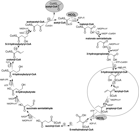
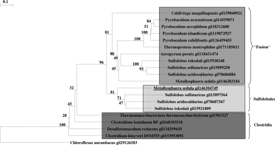
A 3-hydroxypropionate/4-hydroxybutyrate cycle operates in autotrophic CO(2) fixation in various Crenarchaea, as studied in some detail in Metallosphaera sedula. This cycle and the autotrophic 3-hydroxypropionate cycle in Chloroflexus aurantiacus have in common the conversion of acetyl-coenzyme A (CoA) and two bicarbonates via 3-hydroxypropionate to succinyl-CoA. Both cycles require the reductive conversion of 3-hydroxypropionate to propionyl-CoA. In M. sedula the reaction sequence is catalyzed by three enzymes. The first enzyme, 3-hydroxypropionyl-CoA synthetase, catalyzes the CoA- and MgATP-dependent formation of 3-hydroxypropionyl-CoA. The next two enzymes were purified from M. sedula or Sulfolobus tokodaii and studied. 3-Hydroxypropionyl-CoA dehydratase, a member of the enoyl-CoA hydratase family, eliminates water from 3-hydroxypropionyl-CoA to form acryloyl-CoA. Acryloyl-CoA reductase, a member of the zinc-containing alcohol dehydrogenase family, reduces acryloyl-CoA with NADPH to propionyl-CoA. Genes highly similar to the Metallosphaera CoA synthetase, dehydratase, and reductase genes were found in autotrophic members of the Sulfolobales. The encoded enzymes are only distantly related to the respective three enzyme domains of propionyl-CoA synthase from C. aurantiacus, where this trifunctional enzyme catalyzes all three reactions. This indicates that the autotrophic carbon fixation cycles in Chloroflexus and in the Sulfolobales evolved independently and that different genes/enzymes have been recruited in the two lineages that catalyze the same kinds of reactions.
Figures
Similar articles
-
Malonic semialdehyde reductase, succinic semialdehyde reductase, and succinyl-coenzyme A reductase from Metallosphaera sedula: enzymes of the autotrophic 3-hydroxypropionate/4-hydroxybutyrate cycle in Sulfolobales.J Bacteriol. 2009 Oct;191(20):6352-62. doi: 10.1128/JB.00794-09. Epub 2009 Aug 14. J Bacteriol. 2009. PMID: 19684143 Free PMC article.
-
3-Hydroxypropionyl-coenzyme A synthetase from Metallosphaera sedula, an enzyme involved in autotrophic CO2 fixation.J Bacteriol. 2008 Feb;190(4):1383-9. doi: 10.1128/JB.01593-07. Epub 2007 Dec 28. J Bacteriol. 2008. PMID: 18165310 Free PMC article.
-
Convergent Evolution of a Promiscuous 3-Hydroxypropionyl-CoA Dehydratase/Crotonyl-CoA Hydratase in Crenarchaeota and Thaumarchaeota.mSphere. 2021 Jan 20;6(1):e01079-20. doi: 10.1128/mSphere.01079-20. mSphere. 2021. PMID: 33472982 Free PMC article.
-
Unfamiliar metabolic links in the central carbon metabolism.J Biotechnol. 2014 Dec 20;192 Pt B:314-22. doi: 10.1016/j.jbiotec.2014.02.015. Epub 2014 Feb 24. J Biotechnol. 2014. PMID: 24576434 Review.
-
Occurrence, biochemistry and possible biotechnological application of the 3-hydroxypropionate cycle.Appl Microbiol Biotechnol. 2004 Jun;64(5):605-10. doi: 10.1007/s00253-003-1540-z. Epub 2004 Feb 28. Appl Microbiol Biotechnol. 2004. PMID: 14997352 Review.
Cited by
-
Genome-scale reconstruction and analysis of the metabolic network in the hyperthermophilic archaeon Sulfolobus solfataricus.PLoS One. 2012;7(8):e43401. doi: 10.1371/journal.pone.0043401. Epub 2012 Aug 31. PLoS One. 2012. PMID: 22952675 Free PMC article.
-
Identification of missing genes and enzymes for autotrophic carbon fixation in crenarchaeota.J Bacteriol. 2011 Mar;193(5):1201-11. doi: 10.1128/JB.01156-10. Epub 2010 Dec 17. J Bacteriol. 2011. PMID: 21169482 Free PMC article.
-
Screening of metagenomic and genomic libraries reveals three classes of bacterial enzymes that overcome the toxicity of acrylate.PLoS One. 2014 May 21;9(5):e97660. doi: 10.1371/journal.pone.0097660. eCollection 2014. PLoS One. 2014. PMID: 24848004 Free PMC article.
-
Carbon fluxes rewiring in engineered E. coli via reverse tricarboxylic acid cycle pathway under chemolithotrophic condition.J Biol Eng. 2025 Feb 26;19(1):20. doi: 10.1186/s13036-025-00489-w. J Biol Eng. 2025. PMID: 40001153 Free PMC article.
-
The Ruegeria pomeroyi acuI gene has a role in DMSP catabolism and resembles yhdH of E. coli and other bacteria in conferring resistance to acrylate.PLoS One. 2012;7(4):e35947. doi: 10.1371/journal.pone.0035947. Epub 2012 Apr 26. PLoS One. 2012. PMID: 22563425 Free PMC article.
References
-
- Alber, B. E., and G. Fuchs. 2002. Propionyl-coenzyme A synthase from Chloroflexus aurantiacus, a key enzyme of the 3-hydroxypropionate cycle for autotrophic CO2 fixation. J. Biol. Chem. 27712137-12143. - PubMed
-
- Altschul, S. F., W. Gish, W. Miller, E. W. Myers, and D. J. Lipman. 1990. Basic local alignment search tool. J. Mol. Biol. 215403-410. - PubMed
Publication types
MeSH terms
Substances
LinkOut - more resources
Full Text Sources
Other Literature Sources
Molecular Biology Databases
