. 2009 Jul;191(13):4186-94.
doi: 10.1128/JB.01758-08. Epub 2009 May 8.

Two-step assembly dynamics of the Bacillus subtilis divisome

Affiliations

Two-step assembly dynamics of the Bacillus subtilis divisome

Pamela Gamba et al. J Bacteriol. 2009 Jul.

Abstract

Cell division in bacteria is carried out by about a dozen proteins which assemble at midcell and form a complex known as the divisome. To study the dynamics and temporal hierarchy of divisome assembly in Bacillus subtilis, we have examined the in vivo localization pattern of a set of division proteins fused to green fluorescent protein in germinating spores and vegetative cells. Using time series and time-lapse microscopy, we show that the FtsZ ring assembles early and concomitantly with FtsA, ZapA, and EzrA. After a time delay of at least 20% of the cell cycle, a second set of division proteins, including GpsB, FtsL, DivIB, FtsW, Pbp2B, and DivIVA, are recruited to midcell. Together, our data provide in vivo evidence for two-step assembly of the divisome. Interestingly, overproduction of FtsZ advances the temporal assembly of EzrA but not of DivIVA, suggesting that a signal different from that of FtsZ polymerization drives the assembly of late divisome proteins. Microarray analysis shows that FtsZ depletion or overexpression does not significantly alter the transcription of division genes, supporting the hypothesis that cell division in B. subtilis is mainly regulated at the posttranscriptional level.


Figures

FIG. 1.
FIG. 1.
Schematic representation of the B. subtilis divisome. The classical view of the B. subtilis divisome is represented; protein names have been abbreviated by excluding Fts and Div (Z, FtsZ; A, FtsA; L, FtsL; W, FtsW; IB, DivIB; IC, DivIC; IVA, DivIVA).
FIG. 2.
FIG. 2.
Synchronized germination of B. subtilis spores. Spores of EzrA-GFP-, GFP-FtsZ-, FtsW-GFP-, GpsB-GFP-, and DivIVA-GFP-expressing strains were heat shocked and germinated as described in Materials and Methods. (A) Morphological stages of spore germination and outgrowth. Phase-contrast (T0, T60) and fluorescence (membrane staining, Nile Red, T240-300) images of spores are shown (time in minutes). (B to D) Synchrony of the spore preparations was assessed by examining the fraction of phase-bright spores at the early time points (B, average of six independent replicates), the increase in optical density (C, average of six independent replicates), and the appearance of a septal membrane (D, average of two independent replicates). Symbols: ○, GFP-FtsZ; ▴, EzrA-GFP; •, DivIVA-GFP; ⧫, GpsB-GFP; ▪, FtsW-GFP. Error bars indicate standard errors.
FIG. 3.
FIG. 3.
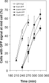
A time delay separates the assembly of FtsZ and EzrA from the recruitment of FtsW, GpsB, and DivIVA. Spores of the five GFP fusion strains were heat shocked and germinated as described in Materials and Methods. GFP pictures were taken every 30 min, starting at 210 min after the beginning of the heat shock. Percentages of cells showing a septal GFP signal were determined for the first round of cell division. At least 150 to 200 cells were analyzed at each time point. The values shown are averages of three replicates from two independent spore preparations. Symbols: ○, GFP-FtsZ; ▴, EzrA-GFP; •, DivIVA-GFP; ⧫, GpsB-GFP; ▪, FtsW-GFP. Error bars indicate standard errors.
FIG. 4.
FIG. 4.
divIVA and ezrA are transcribed immediately after spore germination. Germination of spores of strains with PdivIVA-gfp (•) and PezrA-gfp (▴) was followed by time-lapse microscopy. Images were acquired every 15 min, and the mean GFP fluorescence was recorded. The outgrowth of the spore was set to 0%, while 100% indicates the first appearance of a septum as determined by membrane staining. Trajectories of at least six outgrowing spores of each strain were analyzed, and the highest recorded mean fluorescence value within a single cell trajectory was set to 1 to calculate the relative expression level. Error bars show standard errors. The broken vertical line indicates the time of first appearance of a septum.
FIG. 5.
FIG. 5.
Two clusters of division proteins can be distinguished by time-lapse microscopy. (A and B) Representative cell cycles of EzrA-GFP- and DivIVA-GFP-expressing strains. Spores of EzrA-GFP (A)- and DivIVA-GFP (B)-expressing strains were heat shocked and then germinated on a microscope slide. GFP and FM5-95 pictures were taken every 10 min. The images shown are overlays of both channels. Percentages indicate cell cycle progression, as determined by membrane staining. Asterisks indicate the cells considered in this montage. Arrows indicate the first appearance of EzrA-GFP or DivIVA-GFP at the midcell location. (C and D) Assembly initiation times of eight division proteins (EzrA, FtsZ, FtsA, ZapA, Pbp2B, FtsL, DivIB, and DivIVA) expressed as percentages of cell cycle progression. Growth was followed by time-lapse microscopy as described, and 25 cell cycles were analyzed for each strain. Single-cell values (C) and average values (D) are shown. Error bars indicate standard errors.
FIG. 6.
FIG. 6.
FtsZ overexpression affects the assembly of EzrA but not of DivIVA. (A) Spores of wild-type strains 3312 (ezrA-gfp) and 1803 (divIVA-gfp) and of FtsZ-overexpressing strains PG13 (divIVA-gfp Pxyl-ftsZ) and PG14 (ezrA-gfp Pxyl-ftsZ) were germinated as described in Materials and Methods. The percentage of cells showing a septal GFP signal is shown. Symbols: ▴, EzrA-GFP; ▵, EzrA-GFP (Pxyl-ftsZ); •, DivIVA-GFP; ○, DivIVA-GFP (Pxyl-ftsZ). (B) Representative micrographs of germinating spores of EzrA-GFP and EzrA-GFP (Pxyl-ftsZ) at 210 min after germination. Phase-contrast and GFP channels are shown. Arrows indicate Z rings assembled in short cells immediately after germination.

Similar articles

Cited by

References

    1. Aarsman, M. E., A. Piette, C. Fraipont, T. M. Vinkenvleugel, M. Nguyen-Disteche, and T. den Blaauwen. 2005. Maturation of the Escherichia coli divisome occurs in two steps. Mol. Microbiol. 551631-1645. - PubMed
    1. Anagnostopoulos, C., and J. Spizizen. 1961. Requirements for Transformation in Bacillus subtilis. J. Bacteriol. 81741-746. - PMC - PubMed
    1. Arends, S. J., and D. S. Weiss. 2004. Inhibiting cell division in Escherichia coli has little if any effect on gene expression. J. Bacteriol. 186880-884. - PMC - PubMed
    1. Barák, I., and A. J. Wilkinson. 2007. Division site recognition in Escherichia coli and Bacillus subtilis. FEMS Microbiol. Rev. 31311-326. - PubMed
    1. Bi, E. F., and J. Lutkenhaus. 1991. FtsZ ring structure associated with division in Escherichia coli. Nature 354161-164. - PubMed

Publication types

MeSH terms