. 2009 Jul 10;284(28):18808-15.
doi: 10.1074/jbc.M109.004614. Epub 2009 May 8.

A 20-amino acid module of protein kinase C{epsilon} involved in translocation and selective targeting at cell-cell contacts

Affiliations

A 20-amino acid module of protein kinase C{epsilon} involved in translocation and selective targeting at cell-cell contacts

Barthélémy Diouf et al. J Biol Chem. .

Abstract

In the pituitary gland, activated protein kinase C (PKC) isoforms accumulate either selectively at the cell-cell contact (alpha and epsilon) or at the entire plasma membrane (beta1 and delta). The molecular mechanisms underlying these various subcellular locations are not known. Here, we demonstrate the existence within PKCepsilon of a cell-cell contact targeting sequence (3CTS) that, upon stimulation, is capable of targeting PKCdelta, chimerin-alpha1, and the PKCepsilon C1 domain to the cell-cell contact. We show that this selective targeting of PKCepsilon is lost upon overexpression of 3CTS fused to a (R-Ahx-R)(4) (where Ahx is 6-aminohexanoic acid) vectorization peptide, reflecting a dominant-negative effect of the overexpressed 3CTS on targeting selectivity. 3CTS contains a putative amphipathic alpha-helix, a 14-3-3-binding site, and the Glu-374 amino acid, involved in targeting selectivity. We show that the integrity of the alpha-helix is important for translocation but that 14-3-3 is not involved in targeting selectivity. However, PKCepsilon translocation is increased when PKCepsilon/14-3-3 interaction is abolished, suggesting that phorbol 12-myristate 13-acetate activation may initiate two sets of PKCepsilon functions, those depending on 14-3-3 and those depending on translocation to cell-cell contacts. Thus, 3CTS is involved in the modulation of translocation via its 14-3-3-binding site, in cytoplasmic desequestration via the alpha-helix, and in selective PKCepsilon targeting at the cell-cell contact via Glu-374.


Figures
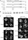
FIGURE 1.
FIGURE 1.
Targeting of PKCβ1, -γ, and -ϵ and their different mutants in GH3B6 cells. Various point mutations were performed in PKCβ1 (GSE mutated into GDE), PKCγ (ADN mutated into AGN) and PKCϵ (GEE mutated into GEA, AEE, or GDE). GH3B6 cells were transiently transfected with the various GFP-tagged constructs (A). All constructs were translated at their expected size (B). Gels were loaded with 15 μg of proteins. Two days after transfection, cells were treated or not with 100 nm PMA for 30 min, and observations were performed with conventional microscopy with an Axiophot 2.0 from Zeiss (C). The scale bars represent 5 μm. Note that targeting is considered to be selective for the cell-cell contact (e.g. wild-type (wt) PKCϵ) when there is no accumulation anywhere else along the cell membrane. The seemingly higher staining detected at the cell-cell contact for PKC isoforms that also translocate along the whole membrane (e.g. wile-type PKCβ1) is because these isoforms are present along both apposed cell membrane constituting the contact. A plot profile is shown in supplemental Fig. 1B to describe a selective versus a nonselective targeting to the cell-cell contact, as we already reported (10).
FIGURE 2.
FIGURE 2.
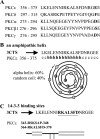
Rationale for the selection of PKCϵ cell-cell contact targeting sequence. A, sequences of PKCϵ, δ, -α, -β1, and -γ upstream of the D(E) amino acid. B, amphipathic α-helix of PKCϵ, and as a comparison, its absence in PKCα. The amphipathic α-helix of PKCϵ is characterized by a hydrophobic residue every 3 or 4 amino acids and by the opposed distribution of the hydrophobic and charged amino acids in the helix. Dark circles represent hydrophobic residues. C, position of the 14-3-3-binding sites.
FIGURE 3.
FIGURE 3.
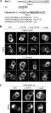
3CTS is a targeting sequence. A, 3CTS bearing or not the substitution of Glu-374 by Gly (mut 3CTS) and a control sequence consisting of GEE plus the 17 amino acids downstream of the GEE (cont) were introduced in PKCδ-GFP, in C1-GFP, and in chimerin-α1-GFP. The PshAI site gacagctgtc in PKCδ was in positions 951–960; the Pm1I site cac/gtg in chimerin-α1 was in positions 406–411. All proteins were expressed at their expected size (supplemental Fig. 3). Two days after transient transfection, GH3B6 cells were observed as in Fig. 1, in the presence or absence of PMA. B, the translocation of PKCδ-GFP (upper) and C1-GFP (lower) with 3CTS, mut 3CTS, or the control sequence is shown. C, the translocation of chimerin-α1-GFP with 3CTS or the control sequence is shown.
FIGURE 4.
FIGURE 4.
Peptide vectorization of 3CTS abolishes selectivity of translocation. RhoB-3CTS and RhoB-scramble 3CTS were chemically linked to the peptide vector sequence Ahx(R-Ahx-R)4. Cells were incubated for 30 min in the presence of either peptide (2.5 μm) followed by a 15-min incubation with 100 nm PMA. A, RhoB fluorescence was observed intracellularly only when the peptides were coupled to the vectorization peptide (left and right). B, in the presence of RhoB-3CTS alone, fluorescence was observed only at the plasma membrane. In these conditions, selectivity was abolished when cells were incubated in the presence of RhoB-3CTS-Ahx(R-Ahx-R)4 (left).
FIGURE 5.
FIGURE 5.
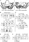
PKCϵ interaction with 14-3-3. A, molecular models of two complexes of a 14-3-3 protein dimer with two 3CTS (left) or with a sequence encompassing Ser-346 and Ser-368 (right) from PKCϵ. The models are derived from the crystal structure Protein Data Bank code 1ib1 of the 14-3-3ζ-serotonin N-acetyltransferase complex (34) using MODELLER 7.0. Left, two identical phosphosites would be predicted to fit perfectly with the crystal structures revealed for various 14-3-3 complexes. The interactions with the 3CTS motif may extend to its helical region including Ile-363. Right, both phosphoserines 346 and 368 would be interacting with the 14-3-3-binding pocket. The predicted helical segment of the central region of 3CTS would bridge the two phosphosites. B, endogenous PKCϵ and PKCϵ-GFP interact with 14-3-3 upon PMA or TRH stimulation. Middle and bottom, effect of the S368A and S346A and of the S346E and S368E mutations on PKCϵ-GFP interaction with 14-3-3. C, C1–3CTS-GFP bearing or not a S368A mutation and 3CTS-GFP bearing or not a S368E or S368A mutation interact with 14-3-3. D and E, incidence of the E374G mutation (D) and of the I363G mutation (E) on the interaction of PKCϵ-GFP or C1–3CTS-GFP with 14-3-3. Experiments shown in B–E have been performed in the presence of 100 nm PMA.
FIGURE 6.
FIGURE 6.
Phosphorylation-dependent interaction of PKCϵ and 3CTS with 14-3-3 after PMA treatment. Extracts (500 μg of proteins) generated from 3CTS-GFP-transfected GH3B6 cells treated or not with 100 nm PMA were used to immunoprecipitate endogenous 3CTS-GFP. 3CTS-GFP bore (B) or not (A) a S368A mutation. Immunoblots were probed with an anti-pSer antibody. 3CTS phosphorylation was increased in the presence of PMA (A) and greatly decreased after substitution of Ser-368 by an alanine (B). Quantification, performed on three independent experiments, is shown at the bottom of each panel. Bars represent S.E. wb, Western blot; WT, wild-type; IP, immunoprecipitation.
FIGURE 7.
FIGURE 7.
Incidence on translocation of S346A and/or S368A or I363G mutation. A, quantification of PKCϵ translocation in TRH-stimulated cells was performed from real-time recordings after transfection with PKCϵ-GFP bearing or not the S368A or the S346A mutation or both and bearing or not the I363G mutation. Results are expressed as percentage (and absolute numbers) of cells translocating or not to the specified location. *, p = 0.04 and **, p = 0.07 against wild-type (wt) PKCϵ-GFP (χ2 test). B, translocation of C1–3CTS-GFP is abolished in the presence of the I363G mutation.
FIGURE 8.
FIGURE 8.
Integrated model for the coordinated involvement of the amphipathic α-helix, the 14-3-3-binding site, and the Glu-374 amino acid of 3CTS in the control of PKCϵ translocation and function. The α-helix is involved very early in the translocation process because its alteration inhibits translocation. Upon PMA or TRH activation, the amount of PKCϵ that localizes at the cell-cell contact might depend on the local amount of 14-3-3 available to bind PKCϵ. The higher the amount of 14-3-3, the lower the amount of PKCϵ translocated to cell-cell contacts will be. This implies that two sets of functions might be regulated by PMA/TRH because both PKCϵ translocation to cell-cell contact and phosphorylations at Ser-368 and Ser-346, which are necessary for PKCϵ/14-3-3 interaction, are PMA/TRH-dependent.

References

    1. Nishizuka Y. (2003) J. Biochem. 133, 155–158 - PubMed
    1. Parker P. J., Murray-Rust J. (2004) J. Cell Sci. 117, 131–132 - PubMed
    1. Lehel C., Olah Z., Jakab G., Anderson W. B. (1995) Proc. Natl. Acad. Sci. U.S.A. 92, 1406–1410 - PMC - PubMed
    1. Schmalz D., Hucho F., Buchner K. (1998) J. Cell Sci. 111, 1823–1830 - PubMed
    1. Schultz A., Ling M., Larsson C. (2004) J. Biol. Chem. 279, 31750–31760 - PubMed

Publication types