. 2009 Jul;37(12):4100-15.
doi: 10.1093/nar/gkp333. Epub 2009 May 8.

Molecular characterization of SMILE as a novel corepressor of nuclear receptors

Affiliations

Molecular characterization of SMILE as a novel corepressor of nuclear receptors

Yuan-Bin Xie et al. Nucleic Acids Res. 2009 Jul.

Abstract

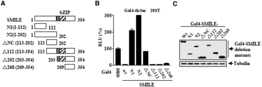
SMILE (small heterodimer partner interacting leucine zipper protein) has been identified as a coregulator in ER signaling. In this study, we have examined the effects of SMILE on other NRs (nuclear receptors). SMILE inhibits GR, CAR and HNF4 alpha-mediated transactivation. Knockdown of SMILE gene expression increases the transactivation of the NRs. SMILE interacts with GR, CAR and HNF4 alpha in vitro and in vivo. SMILE and these NRs colocalize in the nucleus. SMILE binds to the ligand-binding domain or AF2 domain of the NRs. Competitions between SMILE and the coactivators GRIP1 or PGC-1 alpha have been demonstrated in vitro and in vivo. Furthermore, an intrinsic repressive activity of SMILE is observed in Gal4-fusion system, and the intrinsic repressive domain is mapped to the C-terminus of SMILE, spanning residues 203-354. Moreover, SMILE interacts with specific HDACs (histone deacetylases) and SMILE-mediated repression is released by HDAC inhibitor trichostatin A, in a NR-specific manner. Finally, ChIP (chromatin immunoprecipitation) assays reveal that SMILE associates with the NRs on the target gene promoters. Adenoviral overexpression of SMILE represses GR-, CAR- and HNF4 alpha-mediated target gene expression. Overall, these results suggest that SMILE functions as a novel corepressor of NRs via competition with coactivators and the recruitment of HDACs.


Figures

Figure 1.
Figure 1.
SMILE represses GR-, CAR- and HNF4α transactivation. Reporter assays (A–D) were performed as described in the Materials and methods section. HEK293T (293T) cells were cotransfected with 0.1 μg of pcDNA3-HA-GR (A), pcDNA3-HA-CAR (B), pcDNA3-HA-HNF4α (C) or, pcDNA3-HA-PPARγ (D) and 0.1 μg of MMTV-Luc (A), (NR1)5-Luc (B), (HNF4)8-Luc (C) or, PPRE-Luc (D) luciferase reporter vectors, 0.1 μg of pCMV-β-gal as internal control, together with indicated amounts of plasmids expressing wild-type SMILE, SMILE-L (SMILE-83Leu) and SMILE-S (SMILE-1Phe). Twenty-four hours after transfection, the cells were treated with or without 100 nM of dexamethasone (Dex) (A) or rosiglitazone (Rosi) (D) as indicated for 24 h prior to the measurement luciferase activity. The effects of overexpressed SMILE on the protein levels of HA-GR (E) and HA-CAR (F). 293T cells were cotransfected with various plasmids as indicated. Fifty microgram of cellular extracts from the transient transfection assay were subjected to western blot analysis. The proteins of HA-GR, HA-CAR, SMILE and tubulin were detected as described in the Materials and methods section. wt, wild-type.
Figure 2.
Figure 2.
siSMILE increases GR-, CAR- and HNF4α transactivation. (A) Effect of siRNAs for SMILE or SHP on the expression of SMILE and SHP. HepG2 cells were transfected with pSUPER siSMILE-I (siSM#1), siSMILE-II (siSM#2), siSHP or pSUPER [control (con)], and after 72 h the total RNA was isolated. The mRNA levels of SMILE and SHP were measured via RT–PCR analysis, with β-actin shown as a control. The data shown are representative of at least three independent experiments. (B–D) SMILE siRNA induces GR-, CAR- and HNF4α-mediated transactivation in HepG2 cells. HepG2 cells were transfected with pSUPER, pSUPER siSMILE-I (siSM#1), or pSUPER siSMILE-II (siSM#2). After 24 h, the cells were cotransfected with expression vector for GR (B), CAR (C) or HNF4α (D), and MMTV-Luc (B), (NR1)5-Luc (C) or (HNF4)8-Luc (D) luciferase reporter vectors, together with pCMV-β-gal as internal control. Twenty-four hours after transfection, the cells in (B) were treated for 24 h with or without 100 nM Dex prior to the measurement of luciferase activity. The mean and standard deviation (n = 3) of a representative experiment are shown. *P < 0.05; **P < 0.01, using Student's t-test.
Figure 3.
Figure 3.
Interactions and colocalizations of SMILE with NRs. (A–C) In vivo interactions of exogenous GR (A), CAR(B) and HNF4α (C) with exogenous SMILE. 293T cells were cotransfected with expression vectors for HA-GR (A), Flag-mCAR (B), or HA-HNF4α (C) with pEBG-SMILE (GST-SMILE) or pEBG alone (GST). The in vivo GST pull-down assays were performed in the presence or absence of the GR ligand Dex (100 nM) as indicated (A). The complex formation (top panel in A–C, GST puri.) and the amount of HA-GR, Flag-mCAR or HA-HNF4α used for the in vivo binding assay (bottom panel in A–C, Lysate) were determined via western blot using an anti-HA or anti-Flag antibody. The same blot was stripped and reprobed with an anti-GST antibody (middle panel in A–C) to confirm the expression levels of the GST fusion protein (GST-SMILE) and the GST control (GST). In vivo interactions of endogenous GR (D), CAR (E) and HNF4α (F) with endogenous SMILE. Co-immunoprecipitation assays were performed using cell extract from HepG2 cells using indicated antibodies in the presence or absence of 100 nM Dex or 250 nM of TCPOBOP. Endogenous SMILE was immunoprecipitated with GR, CAR and HNF4α (upper panels). The proteins in the cell lysates (middle and lower panels) were analyzed with western blot analysis using indicated antibodies. (G) In vitro GST pull-down assays. Upper panel, 35S-radiolabeled GR protein was incubated with GST, or GST-SMILE fusion proteins in the presence of 100 nM Dex or vehicle (DMSO). Lower panel: 35S-radiolabeled HNF4α, or CAR proteins were incubated with GST, or GST-SMILE fusion proteins. The input lane represents 10% of the total volume of in vitro-translated proteins used for binding assay. Protein interactions were detected via autoradiography. (H–J) Co-localizations of SMILE with NRs. Hela cells grown on coverslips on 12-well plates were transfected with 0.1 μg of expression vectors encoding GFP-SMILE and HA-GR (H), HA-CAR (I) or HA-HNF4α (J). Twelve hours after transfection, the cells (H) were treated with 100 nM Dex for 12 h. For the immunofluorescence of fixed cells, the HA-fusion proteins were detected with dye Alexa 594-conjugated anti-HA monoclonal antibody. The cell images were captured under 400 × magnifications. The data shown are representative of at least three independent experiments.
Figure 4.
Figure 4.
Homodimerization of SMILE is not essential for the repressive function. (A) Structure of human SMILE. The basic region and leucine zipper domains are shown. The leucine zipper mutant SMILE-L (239–267)V indicates the leucine residues between positions 239 and 267 were mutated to valine, as indicated by the arrows. The numbers in the figure indicate the amino-acid residues. (B) In vivo homodimerization possibility analysis of wt SMILE and SMILE-L (239–267)V. 293T cells were cotransfected with expression vectors for Flag-SMILE, Flag-SMILE-L (239–267)V with pEBG-SMILE (GST-SMILE wt) or pEBG-SMILE-L (239–267)V or pEBG alone (GST) as indicated. The complex formation (top panel, GST puri.) and the amount of Flag-SMILE, Flag-SMILE-L (239–267)V used for the in vivo-binding assay (bottom panel, Lysate) were determined by western blot using anti-Flag antibody. The same blot was stripped and reprobed with an anti-GST antibody (middle panel) to verify the expression levels of the GST fusion proteins (GST-SMILE) and the GST control (GST). (C–E) The effect of SMILE-L (239–267)V on GR, CAR and HNF4α-mediated transactivation. HepG2 cells were cotransfected with expression vector for GR (C), CAR (D), or HNF4α (E) and MMTV-Luc (C), (NR1)5-Luc (D), or (HNF4)8-Luc (E) luciferase reporter vectors, pCMV-β-gal as internal control, together with expression vectors for wt SMILE or SMILE-L (239–267)V as indicated. Twenty-four hours after transfection, the cells in (C) were treated with 100 nM Dex for 24 h. Forty-eight hours after tranfection, luciferase activity was measured. wt, wild type. The mean and standard deviation (n = 3) of a representative experiment are shown.
Figure 5.
Figure 5.
Interaction domain of SMILE. (A) Schematic representation of the structures of SMILE mutants. bZIP indicates the basic region leucine zipper domain. The numbers in the figure indicate the amino acid residues. (B–D) In vivo interaction assays between wt SMILE or SMILE mutants and GR (B), CAR (C) or HNF4α (D). 293T cells were cotransfected with expression vectors for HA-GR (B), Flag-CAR (C) or HA-HNF4α (D) with pEBG alone (GST) or pEBG-SMILE (GST-SMILE) fusions as indicated. The in vivo GST pull-down assays in (B) was performed in the presence or absence of GR ligand Dex (100 nM). The complex formation (top panel in B–D, GST puri.) and the amount of HA-GR, Flag-mCAR or HA-HNF4α used for the in vivo-binding assay (bottom panel in B–D, Lysate) were determined by western blot using an anti-HA or anti-Flag antibody. The same blot was stripped and reprobed with an anti-GST antibody (middle panel in B–D) to confirm the expression levels of the GST fusion proteins (GST-SMILE fusions) and the GST control (GST). wt, wild type. The data shown are representative of at least three independent experiments with similar results.
Figure 6.
Figure 6.
SMILE interacts with LBD/AF2 domain of the NRs. (A, C and E) Schematic representation of the structures of the GR (A), CAR (C) and HNF4α (E) mutants. AF1, activation function-1 domain; DBD, DNA-binding domain; LBD, ligand-binding domain; AF2, activation function-2 domain. (B) 35S-radiolabeled GR proteins were incubated with GST, or GST-SMILE fusion proteins in the presence of ligand Dex (100 nM) or vehicle (DMSO). (D and F) 35S-radiolabeled CAR (D) or HNF4α proteins (F) were incubated with GST, or GST-SMILE fusion proteins. The input lane represents 10% of the total volume of in vitro-translated proteins used for the binding assay. Protein interactions were detected via autoradiography. The data shown represent at least three independent experiments with similar results.
Figure 7.
Figure 7.
SMILE competes with coactivators GRIP1 and PGC-1α. Reporter assays in (A–C) were performed as described in the Materials and methods section. The mean and standard deviation (n = 3) of a representative experiment are shown. HepG2 cells were cotransfected with 0.1 μg of indicated reporter plasmids, MMTV-luc (A), (NR1)5-luc (B), or (HNF4)8-Luc (C), and 0.1 μg of pcDNA3-HA-GR (A), pcDNA3-HA-mCAR (B) or pcDNA3-HA-HNF4α (C), together with the indicated quantities of pcDNA3-Flag-SMILE, pSG5-HA-GRIP1 (A) or pcDNA3-HA-PGC-1α (B and C). Twenty-four hours after transfection, the cells were treated with or without GR ligand Dex (100 nM) for 24 h prior to the measurement of luciferase activity. (D) In vitro competition between SMILE and GRIP1 or PGC-1α. 35S-radiolabeled GR (in the presence of 100 nM DEX), or CAR, or HNF4α proteins were incubated with GST, or GST-SMILE fusion proteins, together with an increasing amounts of unlabeled in vitro translated GRIP1 (0, 3, 6 or 12 μl, upper panel) or PGC-1α (0, 3, 6 or 12 μl, middle and lower panel) proteins. After pull-down, the beads were washed and the samples separated on a 12% SDS–PAGE gel and the protein interactions were detected via autoradiography. The data shown represent at least three independent experiments.
Figure 8.
Figure 8.
Intrinsic repressive function of SMILE. (A) Schematic representation of wt SMILE and its deletion mutants fused in-frame to the yeast Gal4 DBD-(1–147 amino acid). bZIP, basic region leucine zipper domain. (B) 293T cells were cotransfected with the reporter plasmid Gal4-tk-Luc and the indicated expression vectors of Gal4-SMILE or Gal4-SMILE deletion mutants together with pCMV-β-gal vector. Forty-eight hours after transfection, the luciferase activity was measured as described in the Materials and methods section. The normalized luciferase activity values are shown as the percentage of the Gal4-tk-Luc reporter activity stimulated by Gal4-DBD. The mean and standard deviation (n = 3) of a representative experiment are shown. (C) Western blot analysis of Gal4-SMILE and Gal4-SMILE deletion mutants. 293T cells were transfected with the indicated Gal-SMILE chimeras. Whole cell extracts (50 μg) were analyzed via western blot using anti-Gal4 rabbit polyclonal antibody.
Figure 9.
Figure 9.
SMILE recruits HDACs. (A–C) TSA releases SMILE-mediated repression on GR and HNF4α. HepG2 cells were cotransfected with 0.1 μg of reporter plasmids, MMTV-luc (A), (HNF4)8-luc (B) or (NR1)5-Luc (C), and 0.1 μg of pcDNA3-HA-GR (A), pcDNA3-HA-HNF4α (B) or pcDNA3-HA-mCAR (C), together with or without 0.2 μg of pcDNA3-Flag-SMILE as indicated. Twenty-four hours after transfection, the cells were treated for 12 h with or without 100 nM of Dex. Then the cells were treated with indicated concentration of TSA for 12 h in the absence or presence of Dex (100 nM). In vivo interactions of HDAC1 (D), HDAC3 (E) and HDAC4 (F) with SMILE. 293T cells were cotransfected with expression vectors for Flag-HDAC1, or HA-HDAC3 or Flag-HDAC4 with pEBG-SMILE (GST-SMILE) or pEBG alone (GST). The complex formation (top panel in D–F, GST puri.) and the amount of Flag- or HA-tagged HDAC fusion proteins used for the in vivo-binding assay (bottom panel in D–F, Lysate) were determined using anti-Flag or anti-HA antibody, respecitively. The same blot was stripped and reprobed with an anti-GST antibody (middle panel in D–F) to confirm the expression levels of the GST fusion protein (GST-SMILE) and the GST control (GST). (G and H) The effect of HDAC siRNAs on the inhibition of GR- and HNF4α-mediated transactivation by SMILE. HepG2 cells were transfected with pSuper siHDAC1, siHDAC3, or siHDAC4 as indicated and after 24 h, the cells were cotransfected with 0.1 μg of indicated reporter plasmids, MMTV-luc (G), or (HNF4)8-Luc (H), and 0.1 μg of pcDNA3-HA-GR (G) or pcDNA3-HA-HNF4α (H), together with 0.1 μg of pcDNA3-Flag-SMILE and after 24 h, the cells were treated with or without 100 nM of Dex for 24 h prior to the measurement of luciferase activity. The mean and standard deviation (n = 3) of a representative experiment are shown. *P < 0.05, using Student's t-test. (I) Effect of siRNAs on the expression of HDACs. HepG2 cells were transfected with pSUPER siHDAC1, siHDAC3, siHDAC4 or pSUPER [control (con)], and after 72 h the total RNA was isolated. The mRNA levels of HDACs were measured via RT–PCR analysis, with β-actin shown as a control. The results are representative of three experiments.
Figure 10.
Figure 10.
SMILE down-regulates the transcription of IGFBP1, CYP2B6 and CYP7A1 gene. (A–C) The recruitment of SMILE on IGFBP-1, CYP2B6 and CYP7A1 promoters is associated with histone deacetlyation. (A and B) HepG2 cells were stimulated with or without 100 nM of Dex (A) or 250 nM of TCPOBOP (B) in the presence or absence of 300 μM TSA as indicated. (C) HepG2 cells were infected with or without adenovirus-HNF4α (Ad-HNF4α) in the presence or absence of 300 μM TSA as indicated. Cell lysates from the treated HepG2 cells were then collected for Chip assays. Chromotian fragments were prepared and immumoprecipitated with the indicated specific antibodies. DNA fragments covering a GRE on IGFBP1 promoter (A, upper panel), or CAR-binding site on CYP2B6 promoter (B, upper panel), BARE-I and BARE-II element on CYP7A1 promoter (C, upper panel) were PCR-amplified as described in the Materials and methods section. The lower panels in A–C indicate the amplification of the control regions. (D–F) Shown are RT–PCR carried out using PCR primers for SMILE, IGFBP1, CYP2B6, CYP7A1, HNF4α and β-actin, using total RNA prepared from HepG2 cells infected with indicated adenovirus vector (Ad-null, Ad-SMILE and Ad-HNF4α) (100 pfu/cell). After 24 h of infection, the cells were stimulated with vehicle (DMSO) or 100 nM of Dex (D) for 2 h or 250 nM of TCPOBOP (E) for 24 h before total RNA was isolated. Data shown are representative of three experiments.

Similar articles

Cited by

References

    1. Lu R, Misra V. Zhangfei: a second cellular protein interacts with herpes simplex virus accessory factor HCF in a manner similar to Luman and VP16. Nucleic Acids Res. 2000;28:2446–2454. - PMC - PubMed
    1. Xie YB, Lee OH, Nedumaran B, Seong HA, Lee KM, Ha H, Lee IK, Yun Y, Choi HS. SMILE, a new orphan nuclear receptor SHP interacting protein, regulates SHP-repressed estrogen receptor transactivation. Biochem. J. 2008;416:463–473. - PubMed
    1. Akhova O, Bainbridge M, Misra V. The neuronal host cell factor-binding protein Zhangfei inhibits herpes simplex virus replication. J. Virol. 2005;79:14708–14718. - PMC - PubMed
    1. Hogan MR, Cockram GP, Lu R. Cooperative interaction of Zhangfei and ATF4 in transactivation of the cyclic AMP response element. FEBS Lett. 2006;580:58–62. - PubMed
    1. Misra V, Rapin N, Akhova O, Bainbridge M, Korchinski P. Zhangfei is a potent and specific inhibitor of the host cell factor-binding transcription factor Luman. J. Biol. Chem. 2005;280:15257–15266. - PubMed

Publication types

MeSH terms