. 2009 Jul;37(12):4116-26.
doi: 10.1093/nar/gkp342. Epub 2009 May 8.

Functional mapping of the interaction between TDP-43 and hnRNP A2 in vivo

Affiliations

Functional mapping of the interaction between TDP-43 and hnRNP A2 in vivo

Andrea D'Ambrogio et al. Nucleic Acids Res. 2009 Jul.

Abstract

Nuclear factor TDP-43 has been reported to play multiple roles in transcription, pre-mRNA splicing, mRNA stability and mRNA transport. From a structural point of view, TDP-43 is a member of the hnRNP protein family whose structure includes two RRM domains flanked by the N-terminus and C-terminal regions. Like many members of this family, the C-terminal region can interact with cellular factors and thus serve to modulate its function. Previously, we have described that TDP-43 binds to several members of the hnRNP A/B family through this region. In this work, we set up a coupled minigene/siRNA cellular system that allows us to obtain in vivo data to address the functional significance of TDP-43-recruited hnRNP complex formation. Using this method, we have finely mapped the interaction between TDP-43 and the hnRNP A2 protein to the region comprised between amino acid residues 321 and 366. Our results provide novel details of protein-protein interactions in splicing regulation. In addition, we provide further insight on TDP-43 functional properties, particularly the lack of effects, as seen with our assays, of the disease-associated mutations that fall within the TDP-43 321-366 region: Q331K, M337V and G348C.


Figures

Figure 1.
Figure 1.
(A) A schematic diagram of the CFTR C155T minigene transfected in our add-back assay (dotted lines represent possible splicing outcomes). (B) Three schematic diagrams of TDP-43 WT, Drosophila TDP-43 (TBPH) and TDP-MUT proteins. The two crosses in the TDP MUT diagram represent the F147L/F149L mutations that make this mutant unable to bind UG repeats. (C) The effect on CFTR exon 9 splicing of adding back these three proteins following knock-down of the endogenous TDP-43. Standard deviations obtained in three independent transfection experiments are shown. The western blots against the endogenous TDP-43, tubulin and FLAG peptide are shown in the lower boxes to show silencing efficiency, equal loading and proper transgene expression.
Figure 2.
Figure 2.
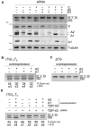
(A) The effect of siRNA knockdown of hnRNP A1, A2 and C on CFTR exon 9 splicing (either singly or in combination). The level of knockdown of each of these hnRNP proteins was assayed by western blot and is reported in the lower panels. The effects of overexpressing the hnRNP A1 and A2 proteins (either alone or in combination) in the presence of TG11T5- and ΔTG-containing CFTR minigenes are shown in (B) and (C), respectively. (D) The effect of overexpressing hnRNP A1, A2 and si-resistant TDP-43 on a CFTR UG11U5 background in the absence of endogenous TDP-43. Standard deviations obtained in three independent transfection experiments are shown for Figure S1B and S1D.
Figure 3.
Figure 3.
(A) A diagram of WT TDP-43 and of two mutants truncated at residues 315 and 366, respectively. The RT–PCRs in (B) show the splicing inhibitory activity of TDP-43 1-366 and 1-315 with respect to TDP WT. Standard deviations obtained in three independent transfection experiments are shown. Western blots against the TDP-43, tubulin and FLAG peptide are shown below to show silencing efficiency, equal expression and proper transgene expression. (C) An EMSA super-shift analysis of the binding between the WT TDP-43, 1-366 and 1-315 TDP-43 mutants with hnRNP A2. Super-shifted complexes are indicated by an arrow.
Figure 4.
Figure 4.
(A) A schematic diagram of the TDP-43 mutants lacking residues 321-366 and the entire C-terminal sequence from residue 216 (ΔC). (B) The splicing inhibitory activity of these two mutants compared to TDP-43 WT. The standard deviations obtained in three independent transfection experiments are reported. Western blots against the TDP-43, tubulin and FLAG peptide are shown below to show silencing efficiency, equal expression and proper transgene expression. (C) An EMSA analysis of the binding between TDP-43 WT, the 321-366 truncation mutant (Δ321-366) and hnRNP A2. Super-shifted complexes are indicated by an arrow.
Figure 5.
Figure 5.
Super-shift EMSA analysis of the binding between TDP-43 WT and hnRNP A2 in the presence of increasing quantities of a peptide (0.5, 1 and 2 μg, respectively, to obtain 60/120/240 molar excess, respectively) spanning residues 321-366 of TDP-43 (p321-366). Equal quantities of a control peptide (pcont) were used as control. Super-shifted complexes are indicated by an arrow.
Figure 6.
Figure 6.
(A) The location of the disease associated missense substitutions found in ALS patients (Q331K, M337V and G348C). Their effect on the ability to bind hnRNP A2 in an EMSA super-shift analysis with hnRNP A2 (indicated by an arrow) and to inhibit splicing compared to TDP-43 WT are reported in (B) and (C), respectively. For the transfection experiments, standard deviations obtained in three independent experiments are shown. Western blots against the TDP-43, tubulin and FLAG peptide are shown below to show silencing efficiency, equal expression and proper transgene expression.
Figure 7.
Figure 7.
The left panel shows the Ponceau protein profile of HeLa cell nuclear extract (NE) and cytoplasmic extract (S100) run on a standard 10% SDS–PAGE gel. Molecular weights (kDa) are shown on the left. The remaining panels show the result of GST-overlay assays performed on a western blot containing the same amount of NE and S100 protein using TDP-43 WT and with TDP-43 carrying the following mutations: Q331K, M337V and G348C. Nuclear and cytoplasmic proteins specifically recognized by the GST-recombinant protein were revealed by ECL following incubation with an anti-GST antibody. The hnRNP cluster recognized by these proteins is indicated.
Figure 8.
Figure 8.
The alignment in (A) shows the sequence homology between the human TDP-43 C-terminal region (hu) and its homologous sequences in Op, Opossum (Monodelphis domestica); Ch, Chicken (Gallus gallus); Fr, Frog (Xenopus laevis); and Ze, Zebrafish (Danio rerio). The 321-366 region is highlighted in bold. (B) The alignment between the full length human TDP-43 and full length Drosophila TBPH. The conserved RRM1 and RRM2 regions are highlighted in bold. (C) An EMSA super-shift analysis with hnRNP A2 of the binding between full length TBPH and a C-terminal truncated TBPH mutant (TBPH ΔC). Super-shifted complexes are indicated by an arrow. (D) A super-shift EMSA analysis of the binding between TBPH and hnRNP A2 in the presence of increasing quantities of the 321-366 TDP-43 peptide (p321-366) and of a control peptide (pcont) in the same quantities described for Figure 5. Super-shifted complexes are indicated by arrows.
Figure 9.
Figure 9.
The left panel shows the Ponceau protein profile of HeLa cell nuclear extract (NE) and cytoplasmic extract (S100) run on a standard 10% SDS–PAGE gel. Molecular weights (kDa) are shown on the left. The remaining panels show the result of three GST-overlay assays using the following proteins: human TDP-43 WT, Drosophila TBPH and Drosophila TBPH ΔC as a control. Nuclear and cytoplasmic proteins specifically recognized by the GST-recombinant protein were revealed by ECL following incubation with an anti-GST antibody. The hnRNP cluster recognized by these proteins is indicated.

References

    1. Buratti E, Baralle FE. Multiple roles of TDP-43 in gene expression, splicing regulation, and human disease. Front. Biosci. 2008;13:867–878. - PubMed
    1. Ayala YM, Misteli T, Baralle FE. TDP-43 regulates retinoblastoma protein phosphorylation through the repression of cyclin-dependent kinase 6 expression. Proc. Natl Acad. Sci. USA. 2008;105:3785–3789. - PMC - PubMed
    1. Wang IF, Wu LS, Chang HY, Shen CK. TDP-43, the signature protein of FTLD-U, is a neuronal activity-responsive factor. J. Neurochem. 2008;105:797–806. - PubMed
    1. Bose JK, Wang IF, Hung L, Tarn WY, Shen CK. TDP-43 overexpression enhances exon 7 inclusion during the survival of motor neuron pre-mRNA splicing. J. Biol. Chem. 2008;283:28852–28859. - PMC - PubMed
    1. Buratti E, Dork T, Zuccato E, Pagani F, Romano M, Baralle FE. Nuclear factor TDP-43 and SR proteins promote in vitro and in vivo CFTR exon 9 skipping. EMBO J. 2001;20:1774–1784. - PMC - PubMed

Publication types

MeSH terms

Substances