. 2009 May 26;106(21):8561-6.
doi: 10.1073/pnas.0812178106. Epub 2009 May 8.

Role of dimerization and substrate exclusion in the regulation of bone morphogenetic protein-1 and mammalian tolloid

Affiliations

Role of dimerization and substrate exclusion in the regulation of bone morphogenetic protein-1 and mammalian tolloid

Richard Berry et al. Proc Natl Acad Sci U S A. .

Abstract

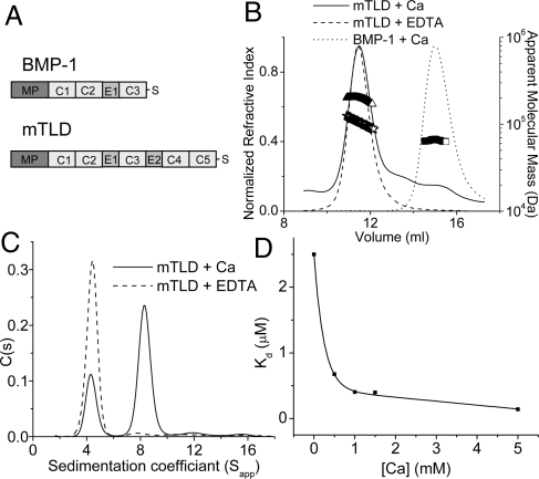
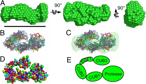
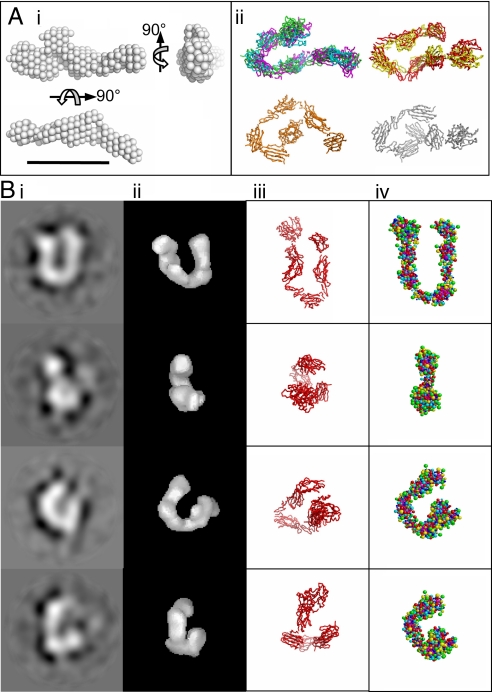
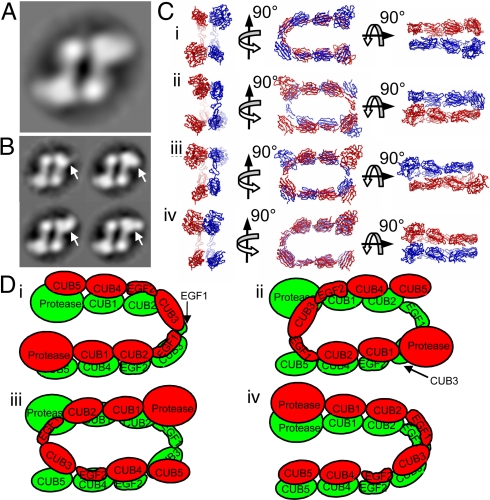
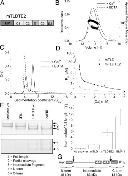
The bone morphogenetic protein (BMP)-1/tolloid metalloproteinases are evolutionarily conserved enzymes that are fundamental to dorsal-ventral patterning and tissue morphogenesis. The lack of knowledge regarding how these proteinases recognize and cleave their substrates represents a major hurdle to understanding tissue assembly and embryonic patterning. Although BMP-1 and mammalian tolloid (mTLD) are splice variants, it is puzzling why BMP-1, which lacks 3 of the 7 noncatalytic domains present in all other family members, is the most effective proteinase. Using a combination of single-particle electron microscopy, small-angle X-ray scattering, and other biophysical measurements in solution, we show that mTLD, but not BMP-1, forms a calcium-ion-dependent dimer under physiological conditions. Using a domain deletion approach, we provide evidence that EGF2, which is absent in BMP-1, is critical to the formation of the dimer. Based on a combination of structural and functional data, we propose that mTLD activity is regulated by a substrate exclusion mechanism. These results provide a mechanistic insight into how alternative splicing of the Bmp1 gene produces 2 proteinases with differing biological activities and have broad implications for regulation of BMP-1/mTLD and related proteinases during BMP signaling and tissue assembly.


Conflict of interest statement

The authors declare no conflict of interest.

Figures

Fig. 1.
Fig. 1.
mTLD, but not BMP-1, forms a calcium-dependent dimer. (A) Domain structure of BMP-1 and mTLD. MP, metalloprotease domain; C, CUB domain; E, EGF-like domain; S, specific region. (B) Summary of MALLS data. In the presence of calcium, mTLD has a molecular mass (triangles) of 196,400 ± 6,284 Da (experimental errors from polydispersity), almost twice that expected for a monomer (100,276 Da). In the presence of EDTA, the apparent molecular mass is reduced to 103,600 ± 2,797 Da (stars). In the presence of calcium, BMP-1 has a molecular mass of 64,920 ± 1,556 Da (squares), similar to that expected for a monomer (70,523 Da). (C) C(s) analysis of mTLD derived from sedimentation velocity analytical ultracentrifugation. (D) The strength of the mTLD self-association was determined at a range of calcium ion concentrations by using sedimentation equilibrium analytical ultracentrifugation.
Fig. 2.
Fig. 2.
Structure of BMP-1. (A) Low resolution ab initio model derived from SAXS data. (B) Six of 8 rigid body models (varying colors) generated by SASREF, also shown superimposed with the ab initio model (C). (D) Representative rigid body model converted into a bead model using SOMO. (E) Proposed BMP-1 structure. (Scale bar, 10 nm; A–D.)
Fig. 3.
Fig. 3.
Structure of the mTLD monomer. (A) (i) Ab initio model generated from SAXS data. (ii) Eight rigid-body models generated by SASREF (different colors) segregated into groups for clarity. (B) Comparison of mTLD structure. (i) Class average images from single-particle image processing (each image is an average of ≈127 particles). (ii) TEM 3D reconstruction calculated by angular reconstitution. (iii) Representative rigid-body SAXS model. (iv) Best-fit bead model. All images in A are to scale. (Scale bar, 10 nm; box size in B, 19.4 nm.)
Fig. 4.
Fig. 4.
Possible models for the mTLD dimer. (A) During TEM, particles are visible in only a single orientation (shown is an average of 851 images). (B) The dataset was reclassified based on the large bulbous region of the right-hand monomer to determine whether this region was flexible. Different conformations are highlighted by white arrows. (C) The mTLD monomer model was used to generate possible models for the dimer. The best fit to AUC measurements was achieved by using a side-by-side nonstaggered arrangement. Within this arrangement, 4 distinct models are possible (shown in ribbon format) with molecules facing the same (C i and iv) or opposite (C ii and iii) directions, and in antiparallel (C i and ii) or parallel (C iii and iv) arrangement. (D) These 4 possible alternatives displayed schematically, with each monomer in a different color. (A and B, box size, 21.6 nm; C is shown on the same scale as A.)
Fig. 5.
Fig. 5.
Oligomerization and chordinase activity of mTLDTE2. (A) Domain structure of mTLDTE2. (B) Summary of MALLS data. The mTLDTE2 has a molecular mass of 106,300 Da (triangles) in the presence of 1 mM calcium ions, and 78,450 Da (squares) in the presence of EDTA. The predicted molecular mass of a monomer is 72,585 Da. (C) Sedimentation velocity AUC of mTLDTE2. (D) The strength of the mTLDTE2 self-association was determined in a range of calcium ion concentrations by using sedimentation equilibrium AUC, and is shown next to that of mTLD. (E) Cleavage of chordin. Purified chordin was incubated in the presence or absence of enzyme as indicated. For clarity, gels were cut into top and bottom layers (shown separately), which were stained to differing extents. (F) Chordinase activity expressed as the mean proportion of the intermediate fragment relative to full-length chordin obtained from 3 independent experiments (error bars represent SEM). (G) Chordin domain structure. CR, cysteine-rich repeat; myc, c-myc epitope; (H)6, hexahistidine tag. Arrows indicated BMP-1 cleavage sites.

Similar articles

Cited by

References

    1. Takahara K, Lyons GE, Greenspan DS. Bone morphogenetic protein-1 and a mammalian tolloid homologue (mTld) are encoded by alternatively spliced transcripts which are differentially expressed in some tissues. J Biol Chem. 1994;269:32572–32578. - PubMed
    1. Pappano WN, Steiglitz BM, Scott IC, Keene DR, Greenspan DS. Use of Bmp1/Tll1 doubly homozygous null mice and proteomics to identify and validate in vivo substrates of bone morphogenetic protein 1/tolloid-like metalloproteinases. Mol Cell Biol. 2003;23:4428–4438. - PMC - PubMed
    1. Hojima Y, van der Rest M, Prockop DJ. Type I procollagen carboxyl-terminal proteinase from chick embryo tendons. Purification and characterization. J Biol Chem. 1985;260:15996–16003. - PubMed
    1. Medeck RJ, Sosa S, Morris N, Oxford JT. BMP-1-mediated proteolytic processing of alternatively spliced isoforms of collagen type XI. Biochem J. 2003;376:361–368. - PMC - PubMed
    1. Unsold C, Pappano WN, Imamura Y, Steiglitz BM, Greenspan DS. Biosynthetic processing of the pro-alpha 1(V)2pro-alpha 2(V) collagen heterotrimer by bone morphogenetic protein-1 and furin-like proprotein convertases. J Biol Chem. 2002;277:5596–5602. - PubMed

Publication types

MeSH terms