. 2009 Jun;136(11):1801-12.
doi: 10.1242/dev.034785.

The neural crest epithelial-mesenchymal transition in 4D: a 'tail' of multiple non-obligatory cellular mechanisms

Affiliations

The neural crest epithelial-mesenchymal transition in 4D: a 'tail' of multiple non-obligatory cellular mechanisms

Jon D Ahlstrom et al. Development. 2009 Jun.

Abstract

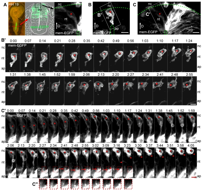
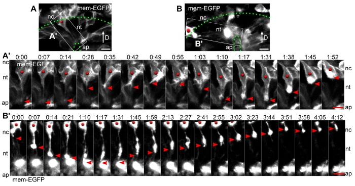
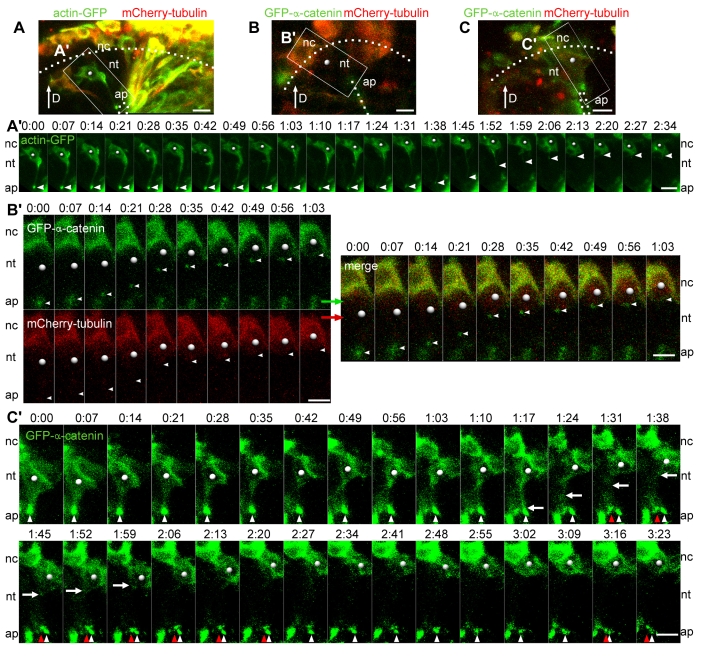
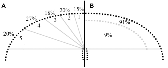
An epithelial-mesenchymal transition (EMT) is the process whereby epithelial cells become mesenchymal cells, and is typified by the generation of neural crest cells from the neuroepithelium of the dorsal neural tube. To investigate the neural crest EMT, we performed live cell confocal time-lapse imaging to determine the sequence of cellular events and the role of cell division in the EMT. It was observed that in most EMTs, the apical cell tail is retracted cleanly from the lumen of the neuroepithelium, followed by movement of the cell body out of the neural tube. However, exceptions to this sequence include the rupture of the neural crest cell tail during retraction (junctional complexes not completely downregulated), or translocation of the cell body away from the apical surface while morphologically rounded up in M phase (no cell tail retraction event). We also noted that cell tail retraction can occur either before or after the redistribution of apical-basolateral epithelial polarity markers. Surprisingly, we discovered that when an EMT was preceded by a mitotic event, the plane of cytokinesis does not predict neural crest cell fate. Moreover, when daughter cells are separated from the adherens junctions by a parallel mitotic cleavage furrow, most re-establish contact with the apical surface. The diversity of cellular mechanisms by which neural crest cells can separate from the neural tube suggests that the EMT program is a complex network of non-linear mechanisms that can occur in multiple orders and combinations to allow neural crest cells to escape from the neuroepithelium.


Figures

Fig. 1.
Fig. 1.
Retraction of the neural crest cell tail occurs most often after complete detachment from the apical surface, but sometimes the cell tail ruptures. (A) A chicken embryo (HH stage 19) electroporated in ovo 18 hours earlier. Imaging was performed on transverse slices at the trunk axial level (red dotted line) to visualize individual cells within the dorsal neural tube (region in black dashed box). (B,C) The mem-EGFP labeled dorsal neural tube cells within the white boxes are shown in the time-lapse series below. The green dotted lines indicate the apical and basal boundaries of the neural tube. (B′) Complete detachment: mem-EGFP labeled cell (red dot) detaches from the apical surface without leaving behind any detectable debris. Time (hours:minutes) is indicated along the top of the series. (C′) Ruptured cell tail: mem-EGFP labeled cell (red dot) ruptures its cell tail (arrowhead), leaving cellular fragments behind (arrow). A more detailed confocal analysis of the cell tail rupture is shown in Fig. S1 in the supplementary material. (C″) Magnified view of the cellular fragment (bright spot) that was part of the cell tail but was left behind during cell tail retraction. Scale bars: 10 μm. D, dorsal; nt, neural tube; nc, neural crest; ap, apical.
Fig. 2.
Fig. 2.
The neural crest EMT can also occur by a non-apical mitosis. (A) The mem-EGFP labeled neural tube cell within the white box is shown in the time-lapse series below. (A′) Non-apical mitosis: mem-EGFP labeled neural tube cell (red dot) rounds up for mitosis, separates from the lumen and divides at a non-apical location. By the end of the time series the daughter cell with the red dot is mostly out of the neural tube. (B) Non-apical mitotic cells are found in uncultured embryos: mem-EGFP labeled embryos were fixed 18 hours after electroporation and cryosectioned. Mitotic cells were detected with an antibody against PH3 (red), the boundaries of the neural tube were visualized with an antibody against α-tubulin (white, labels all cells) and the mem-EGFP signal (green) was used to verify that the non-apical PH3-positive cells within the dorsal neuroepithelium were rounded and not attached to the apical surface. (B′) Magnified view and separate fluorescent channels for the non-apical mitotic cell (*) from the white dashed box in B. Scale bars: 10 μm.
Fig. 3.
Fig. 3.
The behavior of some neural crest cell tails is indecisive. (A,B) The mem-EGFP labeled neural tube cells within the white boxes are shown in the time-lapse series below. (A′) A mem-EGFP labeled neural tube cell (red dot) detaches from the apical surface (arrowhead) and then redeploys a cell process (arrowhead) to the lumen before withdrawing the cell tail completely. (B′) A mem-EGFP labeled neural crest cell (red dot) that is already outside the boundaries of the neural tube sends a cell process to the apical surface (arrowhead) and then retracts it and migrates out of the field of view. Scale bars: 10 μm.
Fig. 4.
Fig. 4.
Time-lapse imaging of dorsal neural tube cells with the circumferential actin belt marker GFP-actin or the adherens junction marker GFP-α-catenin. (A-C) The neural tube cells within the white boxes are shown in the time-lapse series below. (A′) The apical GFP-actin signal (arrowhead) becomes reduced until the cell tail detaches and retracts (arrowhead) from the lumen. (B′) Time-lapse imaging with GFP-α-catenin (green) and mCherry-tubulin (red), showing both the individual and merged channels. When this cell (dot) detaches from the apical surface, the apical GFP-α-catenin signal travels with the end of the cell tail as it retracts (arrowhead). (C′) When this GFP-α-catenin labeled cell (dot) retracts its cell tail (white arrow), the apical GFP-α-catenin signal (white arrowhead) remains behind at the lumen in close proximity to a different apical GFP-α-catenin signal (red arrowhead), not associated with the same cell. A more detailed confocal analysis of the cell tail rupture is shown in Fig. S2 in the supplementary material. Scale bars: 10 μm.
Fig. 5.
Fig. 5.
Time-lapse imaging of dorsal neural tube cells with the apical polarity marker γ-tubulin-GFP (marking the centrosome). (A-C) The neural epithelial cells labeled with γ-tubulin-GFP (green) and mCherry-tubulin (red) within the white boxes are shown in the time-lapse series below. (A′) As this cell undergoes interkinetic nuclear migration and apical mitosis, the centrosome (γ-tubulin-GFP signal) leaves the apical surface and travels basally. Minutes later the nucleus moves towards the apical domain. The nucleus meets the centrosome in a non-apical location, and then both the centrosome and the nucleus travel together to the apical surface, where mitosis occurs. (B′) As this neural tube cell (dot) retracts its tail from the lumen, the γ-tubulin-GFP signal remains at the apical-most tip of the cell tail (white box; corresponds to magnified view below showing γ-tubulin-GFP, mCherry-tubulin and merged channels) as the cell tail is retracted. (C′) In a cell labeled with γ-tubulin-GFP, puncta of γ-tubulin-GFP (arrowheads) are transported from the nucleus towards the apical domain of the epithelium (0-5 hours) until the cell tail detaches from the lumen (5-8 hours). Scale bars: 10 μm.
Fig. 6.
Fig. 6.
The fate of neural tube cells that are separated from the apical surface by a 0°-30° division. (A,B) The mem-EGFP labeled neural tube cells within the white boxes are shown in the time-lapse series below. (A′) A mem-EGFP labeled cell undergoes an apical cell division where the cleavage plane completely separates the basal-most daughter cell (red dot) from the apical surface. The basal-most daughter cell retains a midbody attachment (yellow arrowhead) to the apical daughter cell (yellow dot) for a time, and then the basal-most daughter cell extends a cell process to the lumen (red arrowhead), but retracts this process (red arrowhead) as the cell exits the neural tube. (A″) Confocal z slices from time frame 0:42 show that the basal-most daughter cell (red dot) is completely separated from the apical surface by the plane of cell division. (B′) A mem-EGFP labeled neural tube cell (red dot) undergoes an apical cell division that separates the basal-most daughter cell (red dot) from the apical surface. By the end of this time series, the basal-most daughter cell has clearly re-attached to the lumen (arrows) and remains within the neural tube for the duration of the imaging session. The attachment of the other daughter cell (yellow dot) was out of the confocal viewing field, but the cell body (yellow dot) remains in the neural tube for the duration of the imaging session (not shown). (B″) Confocal z slices of the cell division from time frame 0:21, showing that the basal-most daughter cell (red dot) is completely separated from the apical surface, which can be seen at z=8 μm, even though the dividing cell is bound on either side by mitotic cells at z=1 μm (*) and at z=14 μm (**). Scale bars: 10 μm.
Fig. 7.
Fig. 7.
The surface position and apical-basolateral location of neural tube cell nuclei prior to an EMT. (A) Diagram of the dorsal neural tube showing the percentage of neural crest cells that emigrated from a given region of the neural tube. (B) The percentage of neural tube cell nuclei within a given apical-basal domain 1 hour prior to cell emigration.

References

    1. Ahlstrom, J. D. and Erickson, C. A. (2007). Organization of cells into higher ordered structures. In Principles of Tissue Engineering (ed. R. Lanza, R. Langer and J. P. Vacanti), pp. 67-79. New York: Academic Press.
    1. Baum, B., Settleman, J. and Quinlan, M. P. (2008). Transitions between epithelial and mesenchymal states in development and disease. Semin. Cell Dev. Biol. 19, 294-308. - PubMed
    1. Bilozur, M. E. and Hay, E. D. (1989). Cell migration into neural tube lumen provides evidence for the “fixed cortex” theory of cell motility. Cell Motil. Cytoskeleton 14, 469-484. - PubMed
    1. Bronner-Fraser, M. and Fraser, S. E. (1988). Cell lineage analysis reveals multipotency of some avian neural crest cells. Nature 335, 161-164. - PubMed
    1. Burstyn-Cohen, T. and Kalcheim, C. (2002). Association between the cell cycle and neural crest delamination through specific regulation of G1/S transition. Dev. Cell 3, 383-395. - PubMed

Publication types