. 2009 Aug 1;18(15):2863-74.
doi: 10.1093/hmg/ddp221. Epub 2009 May 9.

Pathogenic mechanisms of tooth agenesis linked to paired domain mutations in human PAX9

Affiliations

Pathogenic mechanisms of tooth agenesis linked to paired domain mutations in human PAX9

Ying Wang et al. Hum Mol Genet. .

Abstract

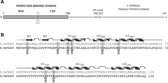
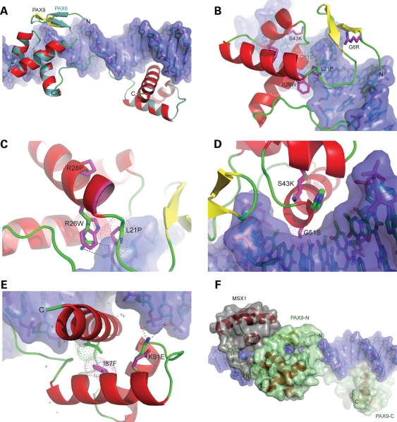
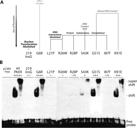
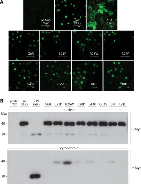
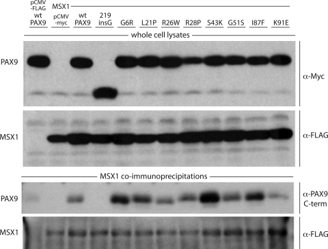
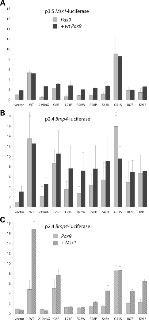
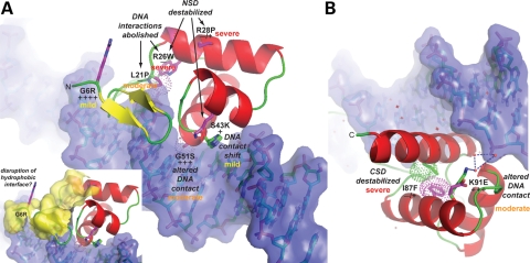
Mutations in the paired-domain transcription factor PAX9 are associated with non-syndromic tooth agenesis that preferentially affects posterior dentition. Of the 18 mutations identified to date, eight are phenotypically well-characterized missense mutations within the DNA-binding paired domain. We determined the structural and functional consequences of these paired domain missense mutations and correlated our findings with the associated dental phenotype variations. In vitro testing included subcellular localization, protein-protein interactions between MSX1 and mutant PAX9 proteins, binding of PAX9 mutants to a DNA consensus site and transcriptional activation from the Pax9 effector promoters Bmp4 and Msx1 with and without MSX1 as co-activator. All mutant PAX9 proteins were localized in the nucleus of transfected cells and physically interacted with MSX1 protein. Three of the mutants retained the ability to bind the consensus paired domain recognition sequence; the others were unable or only partly able to interact with this DNA fragment and also showed a similarly impaired capability for activation of transcription from the Msx1 and Bmp4 promoters. For seven of the eight mutants, the degree of loss of DNA-binding and promoter activation correlated quite well with the severity of the tooth agenesis pattern seen in vivo. One of the mutants however showed neither reduction in DNA-binding nor decrease in transactivation; instead, a loss of responsiveness to synergism with MSX1 in target promoter activation and a dominant negative effect when expressed together with wild-type PAX9 could be observed. Our structure-based studies, which modeled DNA binding and subdomain stability, were able to predict functional consequences quite reliably.


Figures

Figure 1.
Figure 1.
Paired DNA-binding domain of the PAX9 protein. (A) Overall structure of PAX9. The DNA-binding domain of PAX9 is divided into two independent subdomains, an N-terminal (NSD) and a C-terminal (CSD), joined by a short linker segment containing start site (Gly73) of the 219insG PAX9 frameshift mutation. The shift in reading frame produces a truncated polypeptide with a non-native C-terminal segment lacking the second DNA-binding subdomain (CSD), as well as the transcriptional repression (octapeptide motif) and activation domains. (B) Primary and secondary structures of the paired domains of human and mouse PAX6 and PAX9. The DNA-binding subdomains share a homeodomain-like fold, each comprised of three helices. A pair of strands folded into a β-hairpin structure precedes the α-helical homeodomain-like fold of the N-terminal subdomain. Six of the eight missense mutations in the paired domain are within the N-terminal subdomain, three of which are clustered near the N-terminus of the first helix. The secondary structures of the PAX6 crystal structure and PAX9 model (cartoon) were identical. Right angles represent hydrogen-bonded turns in the polypeptide backbone and the grey segment at the N-terminus unstructured residues not included in the crystal structure.
Figure 2.
Figure 2.
Structure-based homology models of PAX9 missense mutant paired domains. (A) Superposition of wild-type PAX9 homology model on the PAX6–DNA complex crystal structure. The α-helical (red) and loop segments (green) of the backbone of the PAX9 model are nearly indistinguishable with those of the PAX6 structural template (turquoise). Note that although the defined secondary structures of model and template are identical (cf. Fig. 1B), the N-terminal β-hairpin of PAX9 (yellow) rendered dissimilarly by the graphics program. (B) Superposition of N-terminal subdomain PAX9 mutant models. The substituted sidechains of each model are depicted (magenta sticks). Similar to the serine sidechain of the PAX6 template, the arginine sidechain of the G6R mutant model extends away from the DNA surface, and without apparent conformational change in the subdomain. (C) Zoomed view of superimposed models of tryptophan and proline mutants in pre-loop and helix of α1 NSD. The molecular surface of the proline sidechain of the L21P mutant (magenta dots) overlaps that of the DNA backbone (violet surface). In the wild-type model, the guanido group of arginine 26 (blue, green stick) forms an ion pair (dashes) with a phosphate group of the DNA backbone that is abrogated by mutation to tryptophan, which is non-isosteric with arginine and alters packing of the subdomain. Introduction of a proline within α1 of the R28P mutant produced a displacement of the N-terminus of the helix (leading edge magenta). (D) Zoomed view of superposition of lysine and serine mutants in α2 and α3 NSD helices. In the S43K mutant model, the sidechain of histidine 50 (imidazole ring, side view) bulges towards the DNA surface to accommodate the lysine sidechain (magenta stick), extended over twice the length of the wild-type serine (green stick). The molecular surface of the serine sidechain of the G51S mutant (magenta dots) protrudes into the surface of the major groove primarily at a cytosine of a G:C base pair. (E) Superposition of C-terminal subdomain PAX9 mutant models. The molecular surface of the phenylalanine sidechain of the I87F mutant (magenta dots) overlaps that of the adjacent isoleucine 126 sidechain in the wild-type subdomain (green dots), resulting in repositioning of the bulky, branched isoleucine sidechain in the mutant (green stick). The wild-type interaction of the ε-amino group of lysine 91 (blue, green stick) with a phosphate group of the DNA backbone through a hydrogen-bonded network of bound water molecules (red spheres) is depicted by dashes. The substituted glutamate sidechain of the mutant model (magenta stick) and positions of additional bound waters (cross-marks) are also shown. (F) Hypothetical superposition model of a ternary complex of PAX9 and MSX1 bound to DNA. The crystal structure of the MSX1–DNA complex (PDB ID: 1IG7) was aligned with the PAX9–DNA model, maintaining the DNA backbones in register and maximizing interaction between the MSX1 and PAX9.
Figure 3.
Figure 3.
Comparison of predicted functional consequences of missense mutations with paired domain consensus site DNA-binding activities. (A) Structure-based homology modeling, ranging from no apparent effect (grey) to severe loss of function (black, bold). The R26W and S43K mutations are predicted to affect both interaction with DNA and protein stability, the G51S mutation DNA interaction only. (B) Electrophoretic mobility shift assay (EMSA) of nuclear extracts from transiently transfected COS7 cells with a paired domain consensus oligonucleotide probe, CD19-2 (A-ins). Anti-myc antibody supershifts (right sample each pair) were performed with the tagged proteins to confirm specificity of labeled duplex oligonucleotide shifts. DNA-binding activities were ranked relative to wild-type PAX9 (+++++) and the 219insG null (–). Weak binding of the R26W and I87F mutant proteins (Table 1, −/+) was detected by prolonged autoradiography. pCMV-myc: nuclear extract from empty vector control transfection.
Figure 4.
Figure 4.
Subcellular immunolocalization of missense mutant proteins. (A) Immunofluorescent detection of Myc-tagged PAX9 proteins in transfected COS7 cells. None of the missense mutations affect nuclear localization, in contrast to the 219insG frameshift that abrogates translocation. (B) Western blot analyses of nuclear and cytoplasmic extracts from transfected COS7 cells with α-Myc antibody. 219insG and small amounts of some missense mutant proteins predicted less stable than wild-type could be detected in the cytoplasmic fractions.
Figure 5.
Figure 5.
Co-immunoprecipitation assay of protein–protein interaction with MSX1. (Upper panels) Western blot analyses of whole cell lysates from COS7 cells cotransfected with Myc-tagged PAX9 proteins and FLAG-tagged MSX1. (Lower panels) Western blot analyses of proteins pulled down with α-FLAG antibody from whole cell lysates. Immunoprecipitated MSX1 was detected with the α-FLAG antibody and co-immunoprecipitated wild-type and missense mutant PAX9 proteins with antibody against the C-terminal transactivation domain of PAX9.
Figure 6.
Figure 6.
Luciferase reporter assays of target promoter upregulation and MSX1 synergy. (A, B) Upregulation of Msx1 and Bmp4 target promoters. Msx1 promoter–reporter (p3.5 Msx1-luciferase) or Bmp4 promoter–reporter (p2.4 Bmp4-luciferase) constructs were co-transfected with mutant Pax9 expression vectors alone (1.5 µg), or in combination with wild-type Pax9 (0.8 µg each). (C) Synergistic upregulation of Bmp4 promoter with MSX1. The Bmp4 promoter–reporter construct was co-transfected with Pax9 expression vector alone (1 µg Pax9, 0.5 µg vector), or in concert with the Msx1 expression vector (1 µg Pax9, 0.5 µg Msx1). Error maxima for wild-type and G51S upregulation in (B) were 20.46 and 25.13 relative units, respectively.
Figure 7.
Figure 7.
Summary of structural and functional consequences of missense mutations and severity of effects on early odontogenesis. (A, B) Zoomed views of the N-terminal (NSD) and C-terminal (CSD) subdomains of PAX9. The structural and functional consequences of each missense mutation, shown schematically in Figure 3A, are indicated along with the observed reduction in in vitro DNA-binding activity relative to wild-type (+++++) as ranked in Figure 3B. In addition, the severity of the effect of each mutation (mild, moderate, severe) on early odontogenesis is indicated by color-coding (yellow, orange, red). The small inset depicts an extensive hydrophobic surface (yellow) that may serve as an interface for recruitment of binding partners to the PAX9:DNA complex. The G6R mutation, which introduces a charged and bulky, albeit relatively flexible sidechain, on the periphery of the hydrophobic patch, could potentially diminish interactions with other proteins within a transcriptional regulatory complex leading to detectable odontogenic affects.

Similar articles

Cited by

References

    1. Thesleff I. Epithelial-mesenchymal signalling regulating tooth morphogenesis. J. Cell Sci. 2003;116:1647–1648. - PubMed
    1. Kapadia H., Mues G., D'souza R. Genes affecting tooth morphogenesis. Orthod. Craniofac. Res. 2007;10:237–244. - PubMed
    1. Peters H., Neubuser A., Balling R. Pax genes and organogenesis: Pax9 meets tooth development. Eur. J. Oral Sci. 1998;106(Suppl. 1):38–43. - PubMed
    1. Peters H., Neubuser A., Kratochwil K., Balling R. Pax9-deficient mice lack pharyngeal pouch derivatives and teeth and exhibit craniofacial and limb abnormalities. Gene Dev. 1998;12:2735–2747. - PMC - PubMed
    1. Mostowska A., Biedziak B., Trzeciak W.H. A novel mutation in PAX9 causes familial form of molar oligodontia. Eur. J. Hum. Genet. 2006;14:173–179. - PubMed

Publication types

MeSH terms

Substances