. 2009 Jul-Aug;6(3):341-50.
doi: 10.4161/rna.6.3.8723. Epub 2009 Jul 14.

A short antisense oligonucleotide masking a unique intronic motif prevents skipping of a critical exon in spinal muscular atrophy

Affiliations

A short antisense oligonucleotide masking a unique intronic motif prevents skipping of a critical exon in spinal muscular atrophy

Natalia N Singh et al. RNA Biol. 2009 Jul-Aug.

Abstract

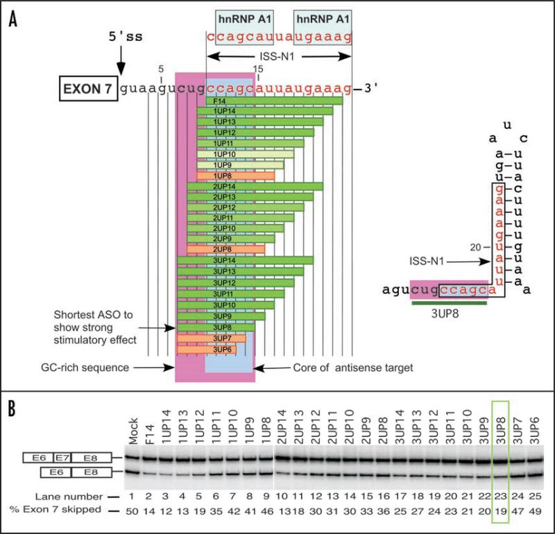
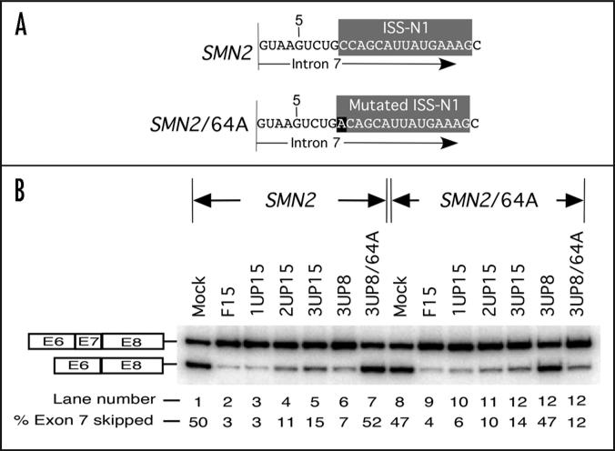
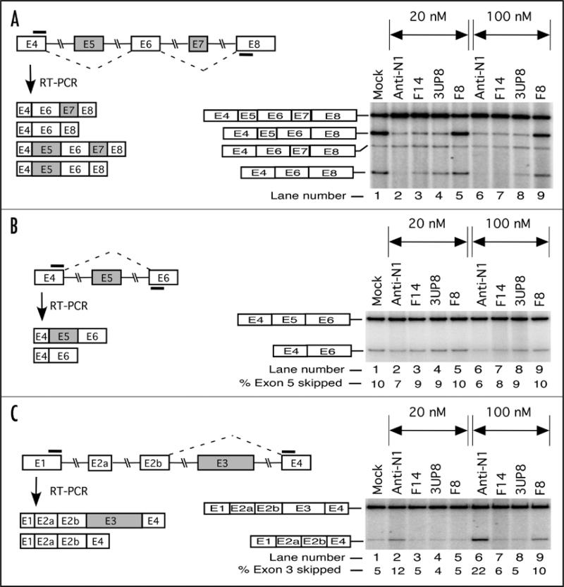
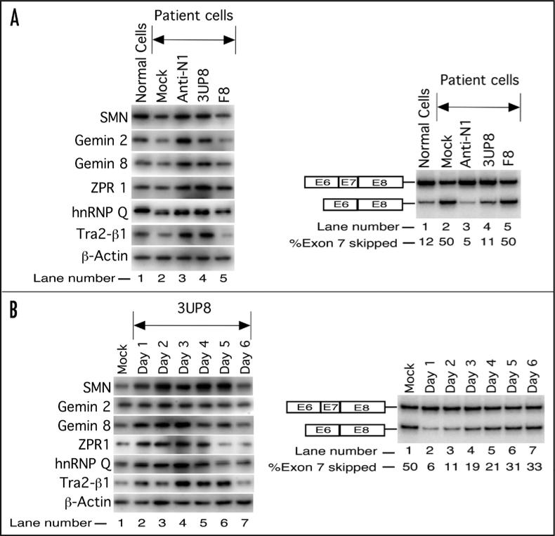
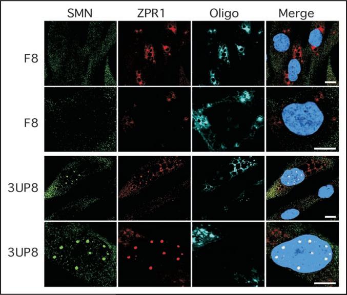
Spinal muscular atrophy (SMA) is the leading genetic cause of infant mortality. Most SMA cases are associated with the low levels of SMN owing to deletion of Survival Motor Neuron 1 (SMN1). SMN2, a nearly identical copy of SMN1, fails to compensate for the loss of SMN1 due to predominant skipping of exon 7. Hence, correction of aberrant splicing of SMN2 exon 7 holds the potential for cure of SMA. Here we report an 8-mer antisense oligonucleotide (ASO) to have a profound stimulatory response on correction of aberrant splicing of SMN2 exon 7 by binding to a unique GC-rich sequence located within intron 7 of SMN2. We confirm that the splicing-switching ability of this short ASO comes with a high degree of specificity and reduced off-target effect compared to larger ASOs targeting the same sequence. We further demonstrate that a single low nanomolar dose of this 8-mer ASO substantially increases the levels of SMN and a host of factors including Gemin 2, Gemin 8, ZPR1, hnRNP Q and Tra2-beta1 known to be down-regulated in SMA. Our findings underscore the advantages and unmatched potential of very short ASOs in splicing modulation in vivo.


Figures

Figure 1
Figure 1
Untra-refined antisense microwalk to identify the shortest stimulatory ASO. (A) Diagrammatic representation of ASOs targeting sequences upstream of ISS-N1. Exon 7 is boxed and the first 24 residues of human SMN intron 7 are shown. Numbering starts from the first position of intron 7. The 5' ss of exon 7 is indicated by a vertical arrow. ASOs blocking different regions are shown as horizontal bars. Sequences of these ASOs are given in Table 1. Boundary of ISS-N1 is demarcated. hnRNP A1 motifs are indicated. Green bars represent ASOs that promote SMN2 exon 7 inclusion. Intensity of green color reflects the strength of stimulatory effect. Tan bars represent ASOs that have no effect on SMN2 exon 7 inclusion. Area highlighted in pink represents the only GC-rich sequence in the first half of human intron 7. Area highlighted in light blue represents the core sequence of the antisense target. Right panel shows the relative positioning of ISS-N1, GC-rich sequence in the context of predicted RNA structure. Green bar represents 3UP8, the shortest ASO to stimulate SMN2 exon 7 inclusion. (B) Splicing pattern of endogenous SMN2 in SMA type I patient fibroblasts (GM03813) treated with different ASOs. Cells were transfected with 20 nM of 2OMePS ASOs and the total RNA for splicing assay was isolated 24 h post transfection. Results were analyzed as described earlier. 3UP8 was the shortest ASO to show stimulatory response (highlighted by green box).
Figure 2
Figure 2
Antisense effect is specific to its target sequence. (A) Diagrammatic representation of intron 7 of SMN2 minigene and its mutant, SMN2/64A. Numbering starts from the first position of human SMN intron 7. ISS-N1 sequence is highlighted in gray. Mutated residue is highlighted in black. (B) Effect of ASOs on splicing of SMN2 minigene and its mutant, SMN2/64A. HeLa cells were transfected with 50 nM of a given ASO and 0.1 μg of minigene in a 24-well plate. Splicing was determined 24 h after transfection. Results were analyzed as described earlier.
Figure 3
Figure 3
Effect of ASOs on alternative splicing of different exons of endogenous SMN2. SMA type I patient fibroblasts (GM03813) were transfected with 20 or 100 nM of selected ASOs in 6-well plates. The total RNA for splicing assay was isolated 24 h post transfection. Spliced products were amplified by RT-PCR with one of the primers being end-labeled. Annealing positions of primers are shown by bars. (A) Left panel depicts the diagrammatic representation of expected spliced products. Right panel shows the results of RT-PCR. Exon 7 skipped, exon 5 skipped and co-excluded products are marked. (B) Left panel depicts the diagrammatic representation of expected spliced products due to skipping of exon 5. Right panel shows the results of RT-PCR. Exon 5 included and exon 5 skipped products are marked. (C) Left panel depicts the diagrammatic representation of expected spliced products due to skipping of exon 3. Right panel shows the results of RT-PCR. Exon 3 included and exon 3 skipped products are marked.
Figure 4
Figure 4
Effect of the shortest stimulatory ASO (3UP8) on levels of cellular proteins in SMA patient cells. (A) Western blot showing the effect of different ASOs. SMA type I patient cells (GM03813) were transfected with 40 nM of selected ASOs and cells were harvested 48 h after transfection. Left panel represents the results of western blot of different proteins, whereas the right panel represents the results of RT-PCR. (B) Time course of 3UP8 effect on the levels of SMN and other factors. SMA type I patient cells (GM03813) were transfected with a single dose of 40 nM of 3UP8 and harvested after every 24 h for six days. Left panel represents the results of western blot of different proteins, whereas the right panel represents the results of RT-PCR.
Figure 5
Figure 5
Confocal images confirming that treatment with short ASO (3UP8) promotes nuclear accumulation of SMN in SMA patient cells. The fibroblasts from SMA type I patient (GM03813) were cultured on coverslips and transfected with 40 nM of F8 (control) or 3UP8 CY3-labeled ASOs. Cells were fixed 48 h after transfection and stained with anti-SMN (Green) and anti-ZPR1 (Red) antibodies. Cells transfected with ASOs were detected by Cy3 fluorescence and presented in pseudo-color (Cyan). DNA was stained with DAPI (blue). The scale bar is 10 mm.

References

    1. Xing Y, Lee C. Relating alternative splicing to proteome complexity and genome evolution. Adv Exp Med Biol. 2007:623:42–9. - PubMed
    1. Nilsen TW. The spliceosome: the most complex macromolecular machine in the cell. Bioessays. 2003;25:1147–9. - PubMed
    1. Matlin AJ, Moore MJ. Spliceosome assembly and composition. Adv Exp Med Biol. 2007;623:14–35. - PubMed
    1. Hertel KJ. Combinatorial control of exon recognition. J Biol Chem. 2008;283:1211–5. - PubMed
    1. Lin S, Fu XD. SR proteins and related factors in alternative splicing. Adv Exp Med Biol. 2007;623:107–22. - PubMed

Publication types

Substances