

doi: 10.1038/nmeth.1329.
Epub 2009 May 10.
TU-tagging: cell type-specific RNA isolation from intact complex tissues
Affiliations
- PMID: 19430475
- PMCID: PMC2783170
- DOI: 10.1038/nmeth.1329
Item in Clipboard
TU-tagging: cell type-specific RNA isolation from intact complex tissues
Nat Methods.
2009 Jun.
Abstract
We found that the combination of spatially restricted uracil phosphoribosyltransferase (UPRT) expression with 4-thiouracil delivery can be used to label and purify cell type-specific RNA from intact complex tissues in Drosophila melanogaster. This method is useful for isolating RNA from cell types that are difficult to isolate by dissection or dissociation methods and should work in many organisms, including mammals and other vertebrates.
Figures
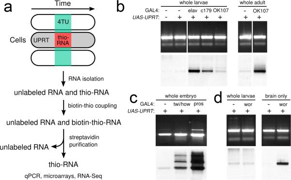
(A) TU-tagging procedure. (B) TU-tagging in larvae, and adults. RNA from larvae expressing GAL4 in no cell type (−), all neurons (elav), muscle cells (c179) or the mushroom body (OK 107) and either with (+) or without (−) UAS-UPRT, and adults of the indicated genotypes were electrophoresed and stained with ethidium bromide to detect all RNA (top) and streptavidinhorseradish peroxidase to detect thio-RNA (bottom). (C) TU-tagging in embryos. 0−16h embryos of the indicated genotypes were treated with 4TU for 2 hours. RNA was analyzed as indicated above. (D). Comparison of RNA labeling before and after tissue isolation. wor, neuroblasts.
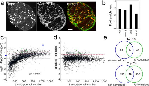
(A) Single confocal section through a brain lobe from a 96h ALH larvae expressing HA:UPRT in all glia (repo-GAL4 UAS-HA:UPRT) stained for HA:UPRT (detected with an HA antibody) and glial nuclei (Repo). Scale bar, 20 μM.(B) fold-enrichment of selected larval glia-specific genes. See supplemental table 1 for all enriched genes. (C) Average microarray ratios from two glia TU-tagging experiments plotted against transcript uracil number. Dashed red line indicates cutoff for top 5% enriched genes. Green dots, previously known larval glia-specific genes; vertical arrowhead, possible false positives; horizontal arrowhead, possible false negatives. (D) TU-tagging microarray ratios after normalization and removal of transcripts with missing UTR annotations (see methods). (E) Comparison of top 1% and 5% enriched genes before and after normalization.
References
Publication types
MeSH terms
Substances
Grants and funding
LinkOut - more resources
Full Text Sources
Other Literature Sources
Molecular Biology Databases
Research Materials
