. 2009 Jun;15(6):649-56.
doi: 10.1038/nm.1958.

Cyclophilin A enhances vascular oxidative stress and the development of angiotensin II-induced aortic aneurysms

Affiliations

Cyclophilin A enhances vascular oxidative stress and the development of angiotensin II-induced aortic aneurysms

Kimio Satoh et al. Nat Med. 2009 Jun.

Abstract

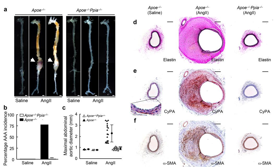
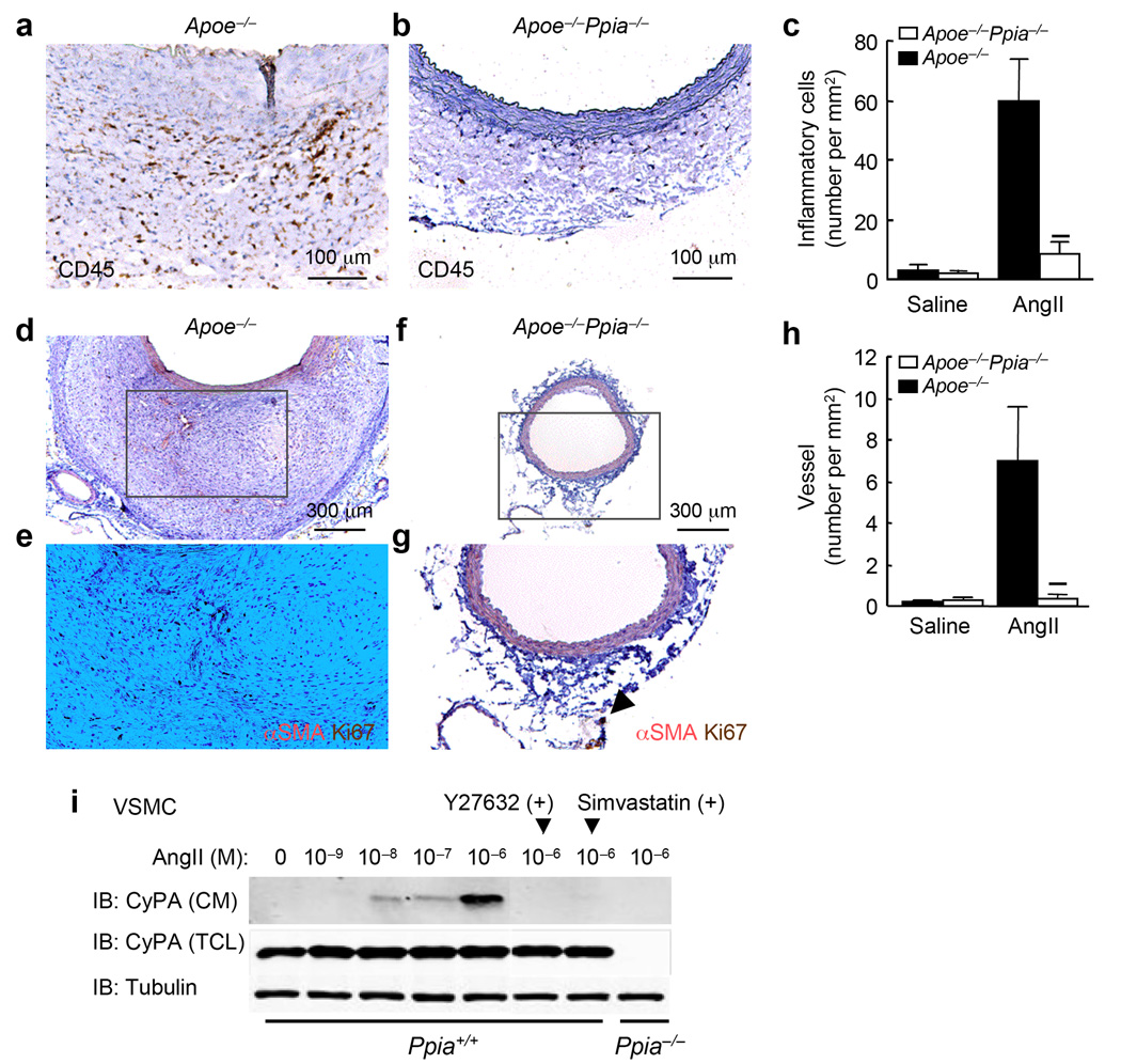
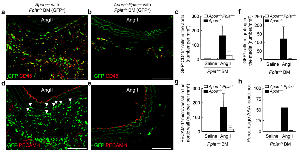
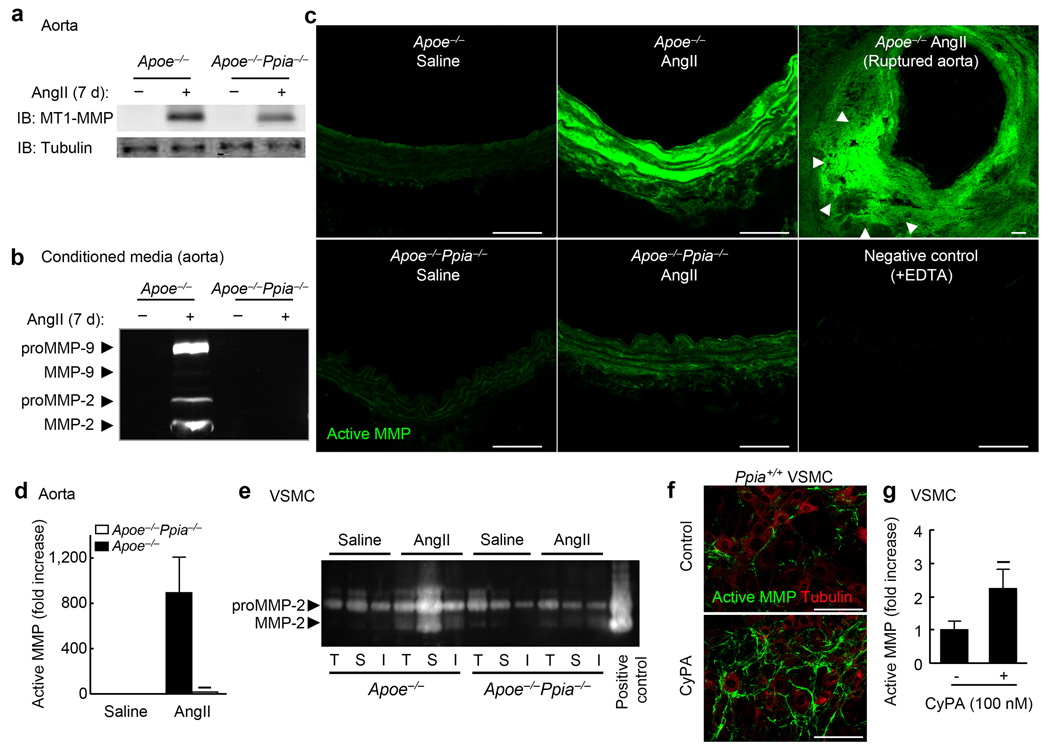
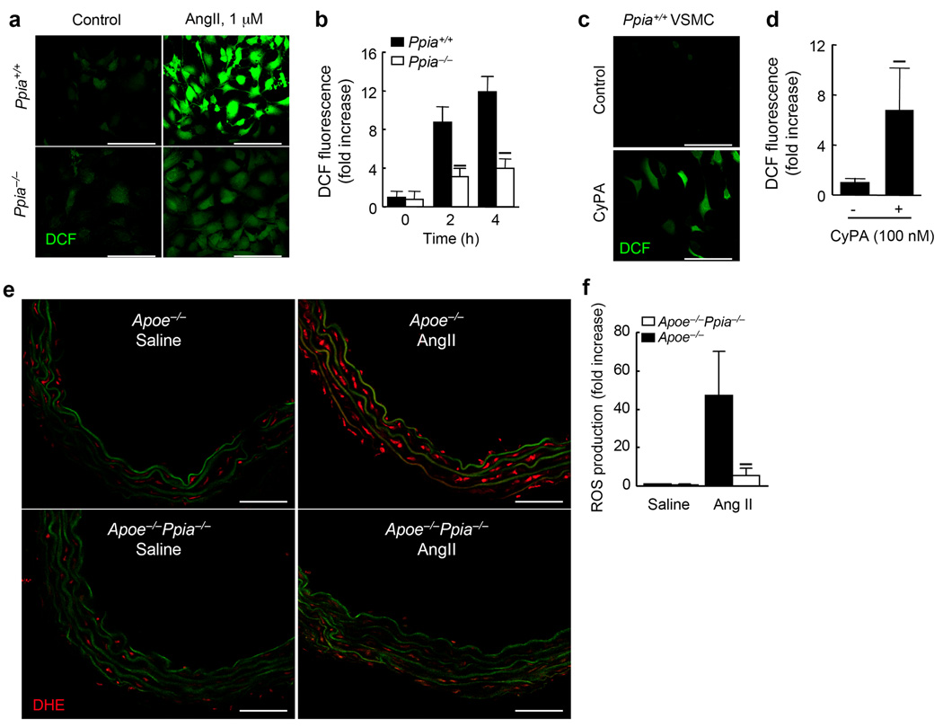
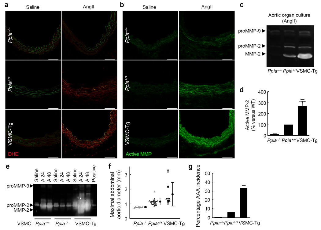
Inflammation and oxidative stress are pathogenic mediators of many diseases, but molecules that could be therapeutic targets remain elusive. Inflammation and matrix degradation in the vasculature are crucial for abdominal aortic aneurysm (AAA) formation. Cyclophilin A (CypA, encoded by Ppia) is highly expressed in vascular smooth muscle cells (VSMCs), is secreted in response to reactive oxygen species (ROS) and promotes inflammation. Using the angiotensin II (AngII)-induced AAA model in Apoe-/- mice, we show that Apoe-/-Ppia-/- mice are completely protected from AngII-induced AAA formation, in contrast to Apoe-/-Ppia+/+ mice. Apoe-/-Ppia-/- mice show decreased inflammatory cytokine expression, elastic lamina degradation and aortic expansion. These features were not altered by reconstitution of bone marrow cells from Ppia+/+ mice. Mechanistic studies showed that VSMC-derived intracellular and extracellular CypA are required for ROS generation and matrix metalloproteinase-2 activation. These data define a previously undescribed role for CypA in AAA formation and suggest CypA as a new target for treating cardiovascular disease.


Figures

Figure 1
Figure 1
CyPA deficiency prevents AngII-induced AAA formation. Apoe−/− and Apoe−/−Ppia−/− mice were infused with AngII or saline for 4 weeks. (a) Representative photographs showing macroscopic features of aneurysms induced by AngII. The arrows indicate typical AAA in Apoe−/− mice. Scale bars, 1 mm. (b) The incidence of AngII-induced AAA was significantly reduced in Apoe−/−Ppia−/− mice (n = 15) compared with Apoe−/− mice (n = 18). There was no AAA formation in the control group (saline infusion) in both Apoe−/− and Apoe−/−Ppia−/− mice (n=4, respectively). (c) Maximal abdominal aortic diameter was significantly reduced in Apoe−/−Ppia−/− mice after AngII infusion for 4 weeks. Triangles represent individual mice; circles represent the mean; error bars denote SD. P < 0.01 compared with AngII-infused Apoe−/− mice. (d) Elastin van Gieson staining of aortic cross-sections of Apoe−/− and Apoe−/−Ppia−/− mice after AngII infusion for 4 weeks. (e, f) The predominant cellular component in the AAA expressing CyPA was VSMC as revealed by immunostaining for CyPA (e) and α-smooth muscle actin (α-SMA) (f), in serial sections. All aortic sections were from the suprarenal aorta. Scale bars, 300 µm.
Figure 2
Figure 2
CyPA deficiency reduces AngII-induced inflammatory cell accumulation and microvessel formation. (a,b) Representative CD45 staining of suprarenal aorta from Apoe−/− and Apoe−/−Ppia−/− mice infused with AngII for 4 weeks. (c) Number of migrating CD45+ cells in the aortic wall in Apoe−/−(n = 9) and Apoe−/−Ppia−/− (n = 7) mice. (dg) Representative immunostaining of α-smooth muscle actin (α-SMA) and Ki67 in suprarenal aorta. (h) Number of proliferating microvessels in the aortic wall. Results are mean ± SD. P < 0.01 compared with Apoe−/− mice. (i) CyPA is secreted from mouse VSMC in response to AngII. Pretreatment with Rho kinase inhibitor Y27632 (30 µM) and simvastatin (30 µM) for 30 min reduced CyPA secretion. CM, conditioned media; TCL, total cell lysate.
Figure 3
Figure 3
Bone marrow (BM) reconstitution shows key role for vascular-derived CyPA in AAA formation. Ppia+/+ BM cells (GFP+) were transplanted into irradiated Apoe−/− or Apoe−/−Ppia−/− mice as described. (a,b) Representative CD45 staining (Alexa Fluor 546, red) of suprarenal aorta from Apoe−/− and Apoe−/−Ppia−/− mice transplanted with Ppia+/+ BM, and infused with AngII for 4 weeks. (c) Number of migrating GFP+CD45+ double-positive cells in the aortic wall in Apoe−/− and Apoe−/−Ppia−/− mice. (d,e) Representative PECAM-1 staining (Alexa Fluor 546, red) of suprarenal aorta from Apoe−/− and Apoe−/−Ppia−/− mice transplanted with Ppia+/+ BM and infused with AngII for 4 weeks. Elastic lamina in the aortic wall demonstrate green auto-fluorescence. Arrows indicate migrating GFP+ cells in the media (d). Number of migrating GFP+ cells in the media (f) or PECAM-1+ microvessels (g) were dramatically higher in the aortic wall of Apoe−/− compared to Apoe−/−Ppia−/− mice. (h) The incidence of AAA in Apoe−/− (n = 9) was much higher than Apoe−/−Ppia−/− (n = 8) mice reconstituted with Ppia+/+ bone marrow after AngII infusion for 4 weeks. Results are mean ± SD. P < 0.01 compared with Apoe−/− mice. Scale bars, 100 µm.
Figure 4
Figure 4
CyPA is crucial for secretion and activation of MMPs. (a) Representative western blot of MT1-MMP expression in mouse aorta after 7 d infusion of AngII. (b) Gelatin zymography for conditioned media from whole aorta organ culture. Aortas from Apoe−/− and Apoe−/−Ppia−/− mice infused with saline or AngII were incubated in media for 20 h. (c) In situ zymography for gelatinase activity. Aortas from Apoe−/− and Apoe−/−Ppia−/− mice infused with saline or AngII for 7 days were analysed. Scale bars, 100 µm. (d) Densitometric analysis of MMP activity (DQ gelatin) changes relative to the density of MMP activity in control Apoe−/− mice (saline-infused). Results are mean ± SD. P < 0.01 compared with Apoe−/− mice. (e) Gelatin zymography for VSMC harvested separately from the thoracic aorta (T), suprarenal aorta (S), and infrarenal aorta (I) of Apoe−/− and Apoe−/−Ppia−/− mice. VSMC from Apoe−/− and Apoe−/−Ppia−/− mice harvested from different portions of aorta were stimulated with AngII (1 µM) for 24 h. (f) Representative in situ zymography (DQ gelatin) of Ppia+/+ VSMC and immunostaining with α-tubulin after stimulation with CyPA (100 nM) for 4 h. (g) Densitometric analysis of MMP activity changes relative to the density of MMP activity in control VSMC. P < 0.01 vs. control VSMC. Results are mean ± SD of 6 independent experiments.
Figure 5
Figure 5
AngII-induced ROS formation in VSMC requires CyPA. (a) Representative DCF staining of mouse aortic VSMC. AngII-induced ROS generation was decreased in CyPA-deficient VSMC. (b) Densitometric analysis of DCF fluorescence in response to AngII shows ~60% reduction in Ppia−/− VSMC at 4 h. Results are mean ± SD of 5 independent experiments. P < 0.01 compared with Ppia+/+ VSMC. (c) Representative DCF staining of Ppia+/+ VSMC in response to 100 nM CyPA. (d) Densitometric analysis of DCF fluorescence in Ppia+/+ VSMC in response to 100 nM CyPA. Results are mean ± SD of 5 independent experiments. P < 0.01 compared with control VSMC. (e) In situ dihydroethidium (DHE) staining of mouse aorta showed decreased DHE staining in Apoe−/−Ppia−/− aortas. Aortas from Apoe−/− and Apoe−/−Ppia−/− mice infused with saline or AngII for 7 d were analysed. Media green fluorescence is from elastin fiber autofluorescence, which appeared both in control and AngII-treated aorta. All sections are shown with the lumen above. Scale bars, 100 µm. (f) Densitometric analysis of DHE fluorescence relative to control Apoe−/− mice (saline-infused). Results are mean ± SD. P < 0.01 compared with Apoe−/− mice.
Figure 6
Figure 6
VSMC-derived CyPA plays a crucial role for aortic ROS production, MMP-2 activation, and AAA formation. (a,b) DHE staining and in situ zymography of supra-renal aorta after treatment with saline or AngII for 7 d. There was increased DHE fluorescence in response to AngII with relative levels: VSMC-Tg > Ppia+/+ > Ppia−/−. All sections are shown with the lumen above. Scale bars, 100 µm. (c) Representative gelatin zymography of conditioned media from mouse aorta after AngII-infusion for 7 d. (d) Active MMP-2 in conditioned media from AngII-treated aortic organ culture shows relative activity: VSMC-Tg > Ppia+/+ > Ppia−/−. P < 0.01 vs. Ppia+/+ aorta. Results are mean ± SD of 3 independent experiments. (e) Representative gelatin zymography of aortic VSMC from Ppia+/+, Ppia−/−, VSMC-Tg mice after treatment with saline, AngII for 24 h (A24), or AngII for 48 h (A48). Positive; MMP-2 positive control. (f) Maximal abdominal aortic diameter significantly increased in VSMC-Tg mice 4 weeks after AngII infusion. Triangles represent individual mice; circles represent the mean; error bars denote SD. (g) The incidence of AngII-induced AAA was significantly increased in VSMC-Tg mice (n = 12) compared with Ppia+/+ mice (n = 17). There was no AAA induction in Ppia−/− mice (n = 8). P < 0.01 compared with AngII-infused Ppia+/+ mice.

References

    1. Kunieda T, et al. Angiotensin II induces premature senescence of vascular smooth muscle cells and accelerates the development of atherosclerosis via a p21-dependent pathway. Circulation. 2006;114:953–960. - PubMed
    1. Griendling KK, FitzGerald GA. Oxidative stress and cardiovascular injury: Part II: animal and human studies. Circulation. 2003;108:2034–2040. - PubMed
    1. Taniyama Y, Griendling KK. Reactive oxygen species in the vasculature: molecular and cellular mechanisms. Hypertension. 2003;42:1075–1081. - PubMed
    1. Bruemmer D, et al. Angiotensin II-accelerated atherosclerosis and aneurysm formation is attenuated in osteopontin-deficient mice. J Clin Invest. 2003;112:1318–1331. - PMC - PubMed
    1. Sun J, et al. Mast cells promote atherosclerosis by releasing proinflammatory cytokines. Nat Med. 2007;13:719–724. - PubMed

Publication types