. 2009;4(5):e5491.
doi: 10.1371/journal.pone.0005491. Epub 2009 May 11.

Involvment of cytosolic and mitochondrial GSK-3beta in mitochondrial dysfunction and neuronal cell death of MPTP/MPP-treated neurons

Affiliations

Involvment of cytosolic and mitochondrial GSK-3beta in mitochondrial dysfunction and neuronal cell death of MPTP/MPP-treated neurons

Agnès Petit-Paitel et al. PLoS One. 2009.

Abstract

Aberrant mitochondrial function appears to play a central role in dopaminergic neuronal loss in Parkinson's disease (PD). 1-methyl-4-phenylpyridinium iodide (MPP(+)), the active metabolite of N-methyl-4-phenyl-1,2,3,6-tetrahydropyridine (MPTP), is a selective inhibitor of mitochondrial complex I and is widely used in rodent and cell models to elicit neurochemical alterations associated with PD. Recent findings suggest that Glycogen Synthase Kinase-3beta (GSK-3beta), a critical activator of neuronal apoptosis, is involved in the dopaminergic cell death. In this study, the role of GSK-3beta in modulating MPP(+)-induced mitochondrial dysfunction and neuronal death was examined in vivo, and in two neuronal cell models namely primary cultured and immortalized neurons. In both cell models, MPTP/MPP(+) treatment caused cell death associated with time- and concentration-dependent activation of GSK-3beta, evidenced by the increased level of the active form of the kinase, i.e. GSK-3beta phosphorylated at tyrosine 216 residue. Using immunocytochemistry and subcellular fractionation techniques, we showed that GSK-3beta partially localized within mitochondria in both neuronal cell models. Moreover, MPP(+) treatment induced a significant decrease of the specific phospho-Tyr216-GSK-3beta labeling in mitochondria concomitantly with an increase into the cytosol. Using two distinct fluorescent probes, we showed that MPP(+) induced cell death through the depolarization of mitochondrial membrane potential. Inhibition of GSK-3beta activity using well-characterized inhibitors, LiCl and kenpaullone, and RNA interference, prevented MPP(+)-induced cell death by blocking mitochondrial membrane potential changes and subsequent caspase-9 and -3 activation. These results indicate that GSK-3beta is a critical mediator of MPTP/MPP(+)-induced neurotoxicity through its ability to regulate mitochondrial functions. Inhibition of GSK-3beta activity might provide protection against mitochondrial stress-induced cell death.


Conflict of interest statement

Competing Interests: The authors have declared that no competing interests exist.

Figures

Figure 1
Figure 1. In vivo and in cellulo GSK-3β activation by MPTP/MPP+.
(A) Seven days after the last saline or MPTP administration, coronal midbrain slices from saline- or MPTP-treated mice were labeled with anti-TH antibody (green) and anti-phospho-Tyr216-GSK-3β (red) as described in Materials and Methods. Merged micrographs illustrated the co-labeling (yellow) of anti-phospho-Tyr216-GSK-3β and anti-TH antibodies. Images are representative of three independent experiments. (B) Saline and MPTP-treated mice were killed one or seven days after the end of the MPTP administration. The brains were homogenized and equal protein amounts were analyzed by Western blotting for phospho-Tyr216-GSK-3β, total GSK-3β and β-tubulin contents. Arrowhead indicates the phospho-Tyr216-GSK-3β, the upper immuno-positive band is a non specific labeling. (C) TSM1 (left) and primary cultures of neurons (right) were incubated with saline (ctrl) or MPP+ (400 µM) for the indicated times (0.5, 1, 2 and 4 h). Total proteins (40 µg) were analyzed for phospho-Tyr216-GSK-3β, phospho-Ser9-GSK-3β, total GSK-3β and β-tubulin by Western blotting. (D) Quantification of blots from three independents experiments performed on TSM1 (left) and primary cultured neurons (right). The phospho-Tyr216-GSK-3β content in every condition was normalized to the total GSK-3β content and expressed as percentage of the control condition. The results are mean±SD and statistical analysis was done using Student t-test. *p<0.01 versus untreated sample.
Figure 2
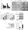
Figure 2. Effect of the down-regulation of GSK-3β activity on MPP+-induced neuronal cell death.
(A) TSM1 and primary cultured neurons were treated with MPP+ (from 0.1 to 2 mM) for 15 h and cell viability was monitored by MTS assay. The data are expressed as the percentage of viable neurons when compared to untreated neurons and are the mean of three independent experiments with triplicate determinations ±S.D. (B) Phase-contrast pictures of representative microscopic fields of TSM1 (left) and primary cultured neurons (right) pre-treated with 25 mM LiCl or 25 µM kenpaullone for 15 min, and then treated with saline or MPP+ for additional 15 h at 37°C. Magnification, ×10. (C) TSM1 neurons (left) and mouse primary neurons (right) were pre-treated with LiCl or kenpaullone (25 mM and 25 µM, respectively), then incubated with MPP+ (400 µM) for 15 h at 37°C and cell viability was assessed using the MTS assay. The data are expressed as the percentage of viable neurons when compared to untreated neurons and are the mean of nine independent experiments with triplicate samples ±S.D. One-way ANOVA *p<0.01 versus saline-treated cells. (D) TSM1 neurons were pre-treated with LiCl (25 mM) for 15 min and co-incubated with 400 µM MPP+ for the indicated times (0.5, 1, 2 and 4 h) and total proteins were extracted and anaylzed for phospho-Tyr216-GSK-3β, total GSK-3β and β-tubulin contents by Western blotting. (E, F) TSM1 cells were transfected with scrambled siRNA (200 nM) or GSK-3β specific siRNA (100 or 200 nM) as described in Materials and Methods. Two days post-transfection, cells lysates were analyzed for total GSK-3β immunoreactivity (upper panel) by Western blotting (F). To determine the correction factor of load, blots were reprobed with the anti-β-tubulin antibody (middle panel). The expression level of GSK-3β was estimated by densitometry analyses performed with a “National Institutes of Health” Image software and expressed as the percent of control conditions. (E) Alternatively, scrambled- or GSK-3β specific- siRNA-transfected cells were treated with 400 µM MPP+ for 15 h at 37°C and the cell viability was monitored using MTS assay. Survival rate in every group was normalized to the untreated control cells. Error bars represent the S.D. for three independent experiments. Statistically significant differences were obtained between siRNA specific GSK-3β- and scrambled siRNA-transfected cells treated with 400 µM MPP+ using Student t-test (*p<0.05).
Figure 3
Figure 3. GSK-3β inhibitors prevented MPP+-induced caspases-9 and -3 activation.
(A, B, C) TSM1 neurons were pre-treated with saline or 25 mM LiCl and then incubated with 400 µM MPP+ for indicated times (1, 2, 4, or 6 h). Intracellular caspase-9 (A) and caspase-3 (B) activities were measured by fluorescence enzymatic assay in cell homogenates. The data are expressed as the percentage of control when compared to untreated neurons and are the mean of three independent experiments with triplicate samples ±S.D. Statistical analysis was done using Student t-test (***p<0.005 versus untreated sample). (C) TSM1 cells were pre-treated or not with 25 mM LiCl and incubated with saline (ctrl) or MPP+ (400 µM) for 1 or 2 h. Total proteins (40 µg) were analyzed for active caspase-3, total caspase-9 and actin contents by Western blotting. (D, E) Primary cultured neurons were pre-treated with saline or 25 mM LiCl and then incubated with 400 µM MPP+ for indicated times (1, 2, 4, or 6 h). Intracellular caspase-9 (D) and caspase-3 (E) activities were measured by fluorescence enzymatic assay in cell homogenates. The data are expressed as the percentage of control when compared to untreated neurons and are the mean of three independent experiments with triplicate samples ±S.D. Statistical analysis was done using Student t-test (*p<0.05 versus untreated sample). Total caspase-9 (D) and active caspase-3 (E) contents were analyzed by Western blotting of primary neurons pre-treated or not with 25 mM LiCl and incubated with saline (ctrl) or MPP+ (400 µM) for 1 or 2 h.
Figure 4
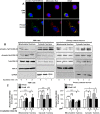
Figure 4. GSK-3β partially localized within mitochondria and MPP+ differentially affected phospho-Tyr216-GSK-3β in mitochondrial and cytosolic fractions.
(A) Fluorescence microscopy pictures of TSM1 and primary cultures of neurons loaded with 100 nM MitoTracker™ (red), then fixed, permeabilized and stained for phospho-Tyr216-GSK-3β (green) and nuclei (DAPI, blue). Magnification ×25. (B, C) TSM1 neurons (left) and cultured primary neurons (right) were treated with 400 µM MPP+ for 1 and 2 h then submitted to subcellular fractionation in order to separate mitochondrial and cytosolic fractions, as described in Materials and Methods. Both fractions were analyzed for phospho-Tyr216-GSK-3β, phospho-Ser9-GSK-3β, total GSK-3β, COXIV and GAPDH contents by Western blotting (B). (C) Densitometry analyses of four to eight independent experiments were carried out to quantify the phospho-Tyr216-GSK-3β content in mitochondrial and cytosolic cellular fractions of vehicle- and LiCl-pretreated TSM1 (left) and primary cultured neurons (right). Data are expressed as percentage of the untreated samples. Statistical analysis was done using Student t-test (* p<0.05, ** p<0.01, ***p<0.005 versus untreated sample).
Figure 5
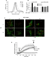
Figure 5. GSK-3β contributed to MPP+-induced TSM1 neuronal cell death through alterations of the mitochondrial membrane potential.
(A) TSM1 neurons were treated with the indicated concentrations of MPP+ for 8 h then cells were stained with JC-1 and analyzed by flow cytometry. (B) TSM1 cells were cultured for 24 h then transfected with scrambled or GSK-3β-specific siRNA (200 nM). Two days post-transfection, cells were pre-treated with saline buffer or 25 mM LiCl or 25 µM kenpaullone (kenp), then co-incubated or not with MPP+ (400 µM) for 8 h. Cells were stained with JC-1 as described in Materials and Methods and prepared for flow cytometry analysis. The results are expressed as the percent of cells with depolarized mitochondrial membrane potential (Ψm). (C, D) intact primary neurons were pre-treated of not with 25 mM LiCl, then co-incubated or not with MPP+ (400 µM) for 4 h. Cells were placed on the stage on a laser-scanning confocal microscope and images were collected every 10 seconds for 10 minutes. One minute after the beginning of experiment, neurons were loaded with 30 nM TMRM (arrow) and one minute before the end of recording, 5 µM FCCP was added to the incubation medium (arrow). (C) Primary neurons pictures were collected 10 minutes after TMRM (green) and Hoechst dye (red) was added to the incubation medium. Magnification, ×63. (D) The fluorescence intensity plots were obtained by selecting eight roi and expressed as the mean±S.D. of the mean gray values derived from all the selected roi in each image.

References

    1. de Lau LM, Breteler MM. Epidemiology of Parkinson's disease. Lancet Neurol. 2006;5:525–535. - PubMed
    1. Schapira AH, Cooper JM, Dexter D, Jenner P, Clark JB, et al. Mitochondrial complex I deficiency in Parkinson's disease. Lancet. 1989;1:1269. - PubMed
    1. Mizuno Y, Ohta S, Tanaka M, Takamiya S, Suzuki K, et al. Deficiencies in complex I subunits of the respiratory chain in Parkinson's disease. Biochem Biophys Res Commun. 1989;163:1450–1455. - PubMed
    1. Parker WD, Jr, Boyson SJ, Parks JK. Abnormalities of the electron transport chain in idiopathic Parkinson's disease. Ann Neurol. 1989;26:719–723. - PubMed
    1. Dauer W, Przedborski S. Parkinson's disease: mechanisms and models. Neuron. 2003;39:889–909. - PubMed

Publication types

MeSH terms

Substances

LinkOut - more resources