

Identification of proteins that interact with podocin using the yeast 2-hybrid system
- PMID: 19430563
- PMCID: PMC2678704
- DOI: 10.3349/ymj.2009.50.2.273
Identification of proteins that interact with podocin using the yeast 2-hybrid system
Abstract
Purpose: As a membrane protein at the insertion site of the slit diaphragm (SD) complex in podocyte foot processes, podocin has been reported to act as a scaffolding protein required to maintain or regulate the structural integrity of the SD. In order to identify proteins that associate or interact with podocin, we screened a mouse kidney complementary DNA (cDNA) library using a yeast 2-hybrid system.
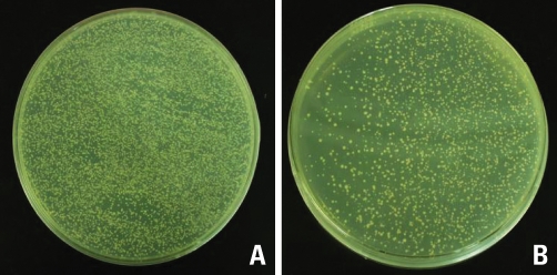
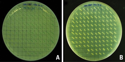
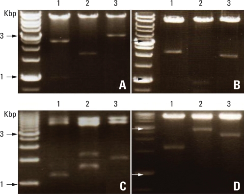
Materials and methods: 1) The full-length cDNA of podocin from the mouse kidney was amplified by Polymerase Chain Reaction (PCR), 2) The PCR product was cloned into a pGBKT7 vector, pGBKT7-podocin, 3) After the pGBKT7-podocin was transformed into AH109, the AH109/pGBKT7-podocin product was obtained, 4) The mouse kidney cDNA library was transformed into the AH109/pGBKT7-podocin and screened by selection steps, 5) Next, twelve clones were cultured and isolated, 6) The yeast-purified plasmids were transformed into Escherichia coli (E. coli) by heat shock, and 7) To identify the activation domain (AD)/library inserts, we digested them with Him III, and the fragments were then sequenced.
Results: 12 positive clones that interacted with podocin were obtained by screening a mouse kidney cDNA library using pGBKT7-podocin. Among them, only 4 clones were found to function at the podocyte where podocin is present.
Conclusion: Additional studies are needed to clarify the role and interaction with podocin and candidates.
Keywords: Podocyte; mouse kidney complementary DNA library; podocin; yeast 2-hybrid system.
Figures


References
-
- Smoyer WE, Mundel P. Regulation of podocyte structure during the development of nephrotic syndrome. J Mol Med. 1998;76:172–183. - PubMed
-
- Somlo S, Mundel P. Getting a foothold in nephrotic syndrome. Nat Genet. 2000;24:333–335. - PubMed
-
- Caulfield JP, Reid JJ, Farquhar MG. Alterations of the glomerular epithelium in acute aminonucleoside nephrosis. Evidence for formation of occluding junctions and epithelial cell detachment. Lab Invest. 1976;34:43–59. - PubMed
Publication types
MeSH terms
Substances
LinkOut - more resources
Full Text Sources
Research Materials
