. 2009 Apr 20:3:5.
doi: 10.3389/neuro.07.005.2009. eCollection 2009.

Right Hemispheric Contributions to Fine Auditory Temporal Discriminations: High-Density Electrical Mapping of the Duration Mismatch Negativity (MMN)

Affiliations

Right Hemispheric Contributions to Fine Auditory Temporal Discriminations: High-Density Electrical Mapping of the Duration Mismatch Negativity (MMN)

Pierfilippo De Sanctis et al. Front Integr Neurosci. .

Abstract

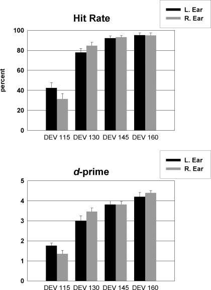
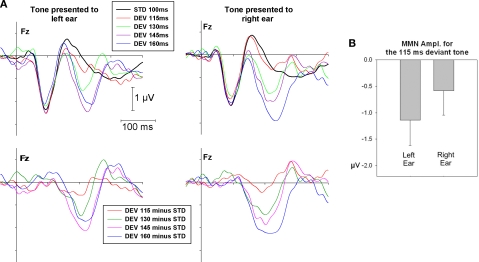
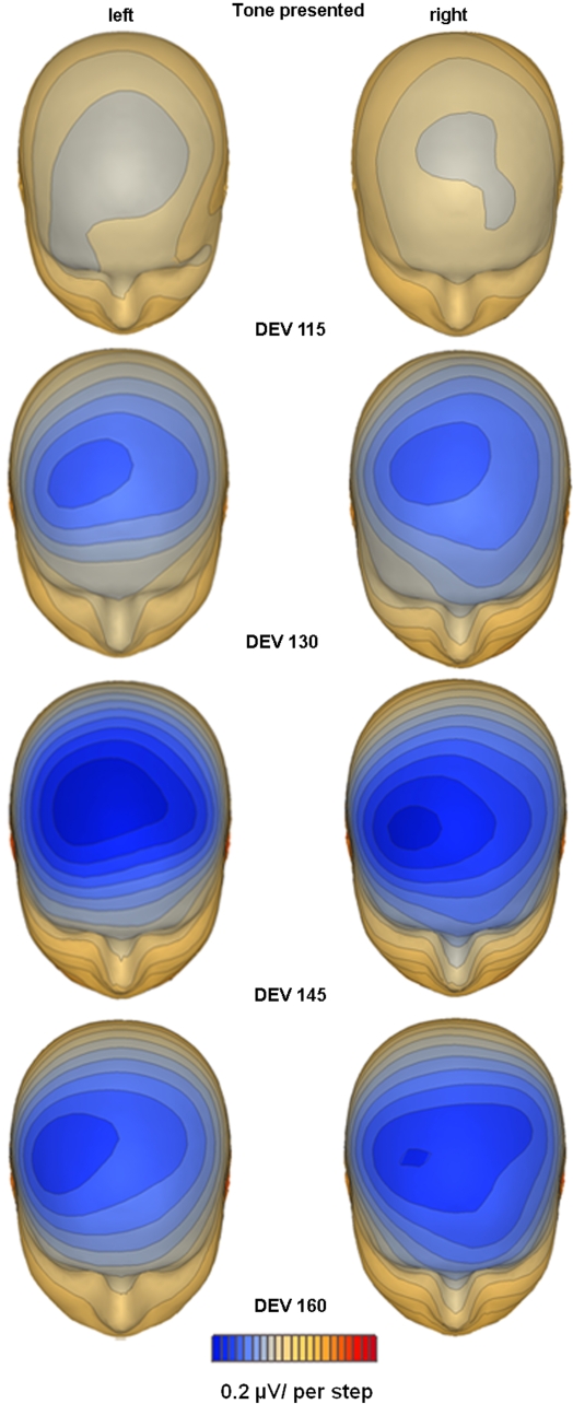
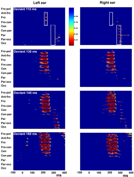
That language processing is primarily a function of the left hemisphere has led to the supposition that auditory temporal discrimination is particularly well-tuned in the left hemisphere, since speech discrimination is thought to rely heavily on the registration of temporal transitions. However, physiological data have not consistently supported this view. Rather, functional imaging studies often show equally strong, if not stronger, contributions from the right hemisphere during temporal processing tasks, suggesting a more complex underlying neural substrate. The mismatch negativity (MMN) component of the human auditory evoked-potential provides a sensitive metric of duration processing in human auditory cortex and lateralization of MMN can be readily assayed when sufficiently dense electrode arrays are employed. Here, the sensitivity of the left and right auditory cortex for temporal processing was measured by recording the MMN to small duration deviants presented to either the left or right ear. We found that duration deviants differing by just 15% (i.e. rare 115 ms tones presented in a stream of 100 ms tones) elicited a significant MMN for tones presented to the left ear (biasing the right hemisphere). However, deviants presented to the right ear elicited no detectable MMN for this separation. Further, participants detected significantly more duration deviants and committed fewer false alarms for tones presented to the left ear during a subsequent psychophysical testing session. In contrast to the prevalent model, these results point to equivalent if not greater right hemisphere contributions to temporal processing of small duration changes.

Keywords: auditory temporal resolution; event-related potentials; hemispheric asymmetry.


Figures

Figure 1
Figure 1
Percent Hit-rates (top panel) and d-prime values (bottom panel) for each degree of temporal deviant and ear of delivery in the behavioral task.
Figure 2
Figure 2
(A) Grand mean ERPs at Fz for standard and deviant tones of 115, 130, 145 and 160 ms duration presented to the left and right ear (top panel) as well as MMN response (bottom panel) as subtraction waveform (deviant − standard). (B) Average MMN amplitude and standard error for the 115 ms deviant presented to the left and right ear.
Figure 3
Figure 3
Topographic mapping of spline-interpolated potential distribution representing the MMN response as subtraction waveform (deviant – standard). The MMN is plotted at peak latency for each ear of delivery and degree of deviance separately. A distinct lateralization of the duration MMN focus over the right hemi-scalp is evident in each condition, except for the condition where no significant MMN activation was evident, e.g. the smallest degree of deviance present to the right ear.
Figure 4
Figure 4
Statistical cluster plots for degree of duration deviants of 115, 130, 145 and 160 ms presented to the left ear (left panel) and right ear (right panel). Color values indicate the result of pointwise t-tests evaluating differences between standard-tone and deviant-tone ERPs across time (x-axis) and electrode positions (y-axis) for the entire 128-electrode montage. For clarity, only p-values <0.05 are color encoded. Fro-pol, Fronto-polar; Ant-fro, Anterior-frontal, Fro, Frontal; Fro-cen, Fronto-central; Cen, Central; Cen-par, Central-parietal; Par, Parietal; Par-occ, Parietal-occipital; Occ, Occipital.
Figure 5
Figure 5
The dipolar solution of the MMN component with sources bilateral in the superior temporal gyri and right inferior frontal gyrus. Dipoles are shown in the Talairach-transformed brain of an individual subject. Color dots by the graphs illustrating mean dipole strength indicate the corresponding color-matched dipole. For each dipole and degree of deviation source strength was calculated as mean of the corresponding source waveform in a time window from 190 to 240 ms. Mean strength of dipole in the left and right STG increased with degree of deviation. No significant change was observed for the right IFG dipole.

References

    1. Abramson A. S. (1977). Laryngeal timing in consonant distinctions. Phonetica 34, 295–303 - PubMed
    1. Amenedo E., Escera C. (2000). The accuracy of sound duration representation in the human brain determines the accuracy of behavioral perception. Eur. J. Neurosci. 12, 2570–257410.1046/j.1460-9568.2000.00114.x - DOI - PubMed
    1. Belin P., McAdams S., Thivard L., Smith B., Savel S., Zilbovicius M., Samson S., Samson Y. (2002). The neuroanatomical substrate of sound duration discrimination. Neuropsychologia 40, 1956–196410.1016/S0028-3932(02)00062-3 - DOI - PubMed
    1. Boemio A., Fromm S., Braun A., Poeppel D. (2005). Hierarchical and asymmetric temporal sensitivity in human auditory cortices. Nat Neurosci. 8, 389–39510.1038/nn1409 - DOI - PubMed
    1. Brown C. P., Fitch R. H., Tallal P. (1999). Sex and hemispheric differences for rapid auditory processing in normal adults. Laterality 4, 39–50 - PubMed

LinkOut - more resources