Review
. 2009 Mar;104(2):320-44.
doi: 10.1590/s0074-02762009000200029.

Why prevent, diagnose and treat congenital toxoplasmosis?

Affiliations
Review

Why prevent, diagnose and treat congenital toxoplasmosis?

Rima McLeod et al. Mem Inst Oswaldo Cruz. 2009 Mar.

Abstract

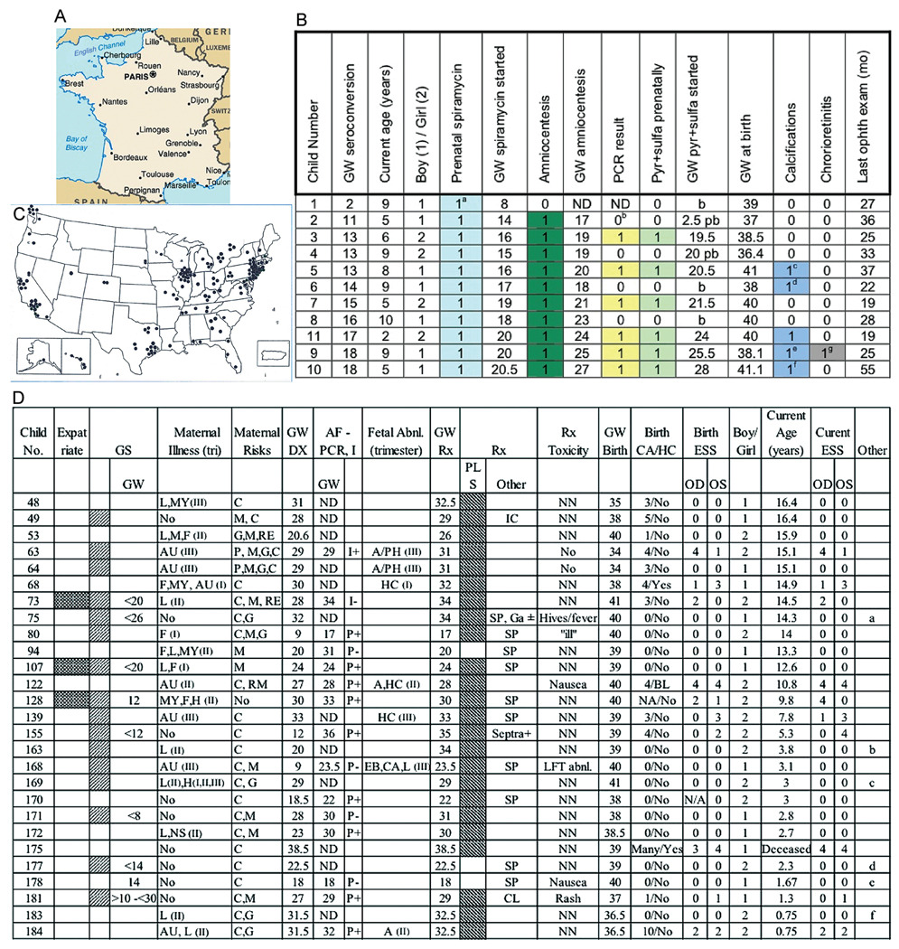
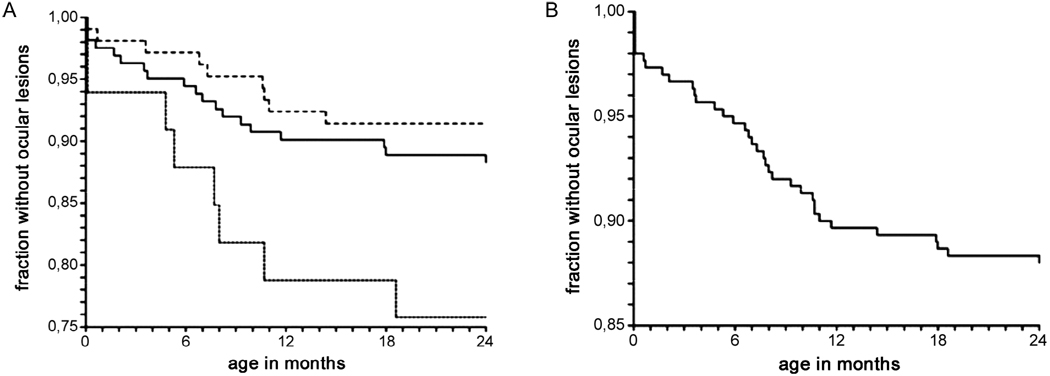
Evidence that prevention, diagnosis and treatment of toxoplasmosis is beneficial developed as follows: anti-parasitic agents abrogate Toxoplasma gondii tachyzoite growth, preventing destruction of infected, cultured, mammalian cells and cure active infections in experimental animals, including primates. They treat active infections in persons who are immune-compromised, limit destruction of retina by replicating parasites and thereby treat ocular toxoplasmosis and treat active infection in the fetus and infant. Outcomes of untreated congenital toxoplasmosis include adverse ocular and neurologic sequelae described in different countries and decades. Better outcomes are associated with treatment of infected infants throughout their first year of life. Shorter intervals between diagnosis and treatment in utero improve outcomes. A French approach for diagnosis and treatment of congenital toxoplasmosis in the fetus and infant can prevent toxoplasmosis and limit adverse sequelae. In addition, new data demonstrate that this French approach results in favorable outcomes with some early gestation infections. A standardized approach to diagnosis and treatment during gestation has not yet been applied generally in the USA. Nonetheless, a small, similar experience confirms that this French approach is feasible, safe, and results in favorable outcomes in the National Collaborative Chicago-based Congenital Toxoplasmosis Study cohort. Prompt diagnosis, prevention and treatment reduce adverse sequelae of congenital toxoplasmosis.


Figures

Fig. 1
Fig. 1
A: Toxoplasma gondii tachyzoites in tissue culture with medium alone; B: with antimicrobial agent. Note growth of parasite and destruction of host cells (adapted from Zuther et al. 1999, with permission).
Fig. 2
Fig. 2
A: resolution of hydrocephalus and brain growth following treatment and shunt in child with congenital toxoplasmosis (Swisher et al. 1994, with permission); B: resolution or diminution of size of intracerebral calcifications during treatment for congenital toxoplasmosis in the first year of life. Cranial CT scans were obtained in the neonatal period and at one year of age. Each cranial CT scan was reviewed by the same study neuroradiologist. Calcification size and number were computed (Patel et al. 1996, with permission). Thirty two (82%) of 39 children had calcifications that diminished or resolved and seven (18%) had calcifications that remained the same size (C) and appearance of brain abscesses in a patient with a cardiac transplant (Ryning et al. 1979, with permission) (D) and a patient with toxoplasmic encephalitis who had AIDS in the pre-HAART era (Levin et al. 1983, with permission). Lesions resolved and clinical status improved for both the patients shown in C and D when they were treated with pyrimethamine and sulfadiazine; E: active retinal lesion before (left) and within a month of initiating treatment (right).
Fig. 3
Fig. 3
A: improved outcomes in treated children contrasted with earlier cohorts and comparing a higher and lower dose regimen in a randomized controlled trial. Frequency of outcomes for each endpoint for patients in our study, compared with the frequency in the literature (Eichenwald 1960, Saxon et al. 1973, Wilson et al. 1980, Koppe et al. 1982, 1986, McLeod et al. 2006a). There is no visible trend for superiority or statistically significant superiority for treatment arm 1 or treatment arm 2 at this time. Results may differ in the future, because the majority of the children enrolled are entering adolescence and early adulthood, a critical time when outcomes may vary. It is also important that outcomes of offspring of the treated children be established in the following years of this study. 1: treatment arm 1 [daily doses of pyrimethamine (1 mg/kg) for two months]; 2: treatment arm 2 [daily doses of pyrimethamine (1 mg/kg) for six months. Following daily dosing with pyrimethamine, this dose was administered on each Monday, Wednesday and Friday. 50 mg/kg sulfadiazine was administered throughout as was calcium leukovorin]; B: Kaplan-Meier Curves showing the outcomes for each endpoint for patients in the pooled feasibility/observational phase and the randomized phase who received treatment 1 (solid line) or treatment 2 (dotted line). There is no visible trend for superiority or statistically significant superiority at this time. Results may differ in the future, because the majority of the children enrolled are entering adolescence and early adulthood, a critical time when outcomes may vary. It is also important that outcomes of offspring of the treated children be established in the following years of this study. IQ: intelligence quotient; Rx1: treatment arm 1; Rx2: treatment arm 2 (adapted from McLeod et al. 2006a, with permission.)
Fig. 4
Fig. 4
A: Koppe study visual outcome for 12 children who were asymptomatic at birth, untreated or treated less than once a month and evaluated when they were six and 20 years old. Percentage of children with retinal disease. Adverse outcomes in untreated congenital toxoplasmosis or when congenital toxoplasmosis was treated for only one month; B: Eichenwald study outcome for 101 patients at ≥ 4 years old. Asterisk: patients with neurologic disease had otherwise undiagnosed central nervous system disease in the first year of life (n = 70); †: patients with generalized disease had otherwise undiagnosed non-neurologic disease in the first two months of life (n = 31); C: children in the NCCCTS in the moderate/severe categories had as severe disease as did children in the Eichenwald series (data are from Eichenwald 1960, Koppe et al. 1982, , Labadie & Hazeman 1984, McLeod et al. 2006a, with permission); D: new eye lesions in children who missed treatment in the first year of life; E: new eye lesions in children treated in the first year of life. In D and E, incidence rate: # of patients with new lesions per person-year. Blue shaded area in D and E is confidence interval.
Fig. 5
Fig. 5
A–F: association of presence of hydrocephalous “shown in brain computed tomography and magnetic resonance image of brains of congenitally infected children” (A, B) and brain at pathologic examination showing characteristic periacqueductal inflammation and necrosis (C) with (D) presence of the HLA DQ3 gene in congenitally infected infants and mothers of the children (from Mack et al. 1999, with permission); E: retinal scars; F: association of alleles of Col2A and ABC4r with hydrocephalus and chorioretinal disease in toxoplasmosis and diagnosis; LT: left eye disease; RT: right eye, representative of eye disease (data adapted from Jamiesen et al. 2008, with permission).
Fig. 6
Fig. 6
French approach to diagnosis and treatment of congenital toxoplasmosisin earlier decades and outcomes using this approach (from Boyer et al. 2008). a: from Desmonts & Courveur 1974a; b: from Hohlfeld et al. 1994a; c: from Daffos et al. 1988; d: from Hohlfeld et al. 1989, Brézin et al. 1993.
Fig. 7
Fig. 7
parasite isolation, treatment and outcome in congenital toxoplasmosis. A: reduction in isolation from placenta of infected infants following treatment with spiramycin, pyrimethamine/sulfadiazine versus (B) no treatment. Amniotic fluid (AF) parasite burden predicts severity of disease in congenital toxoplasmosis. Correlation between Toxoplasma concentrations in AF samples and gestational age at maternal infection for the 86 cases. Severity of the infection is represented in each case by ■ if severe signs of infection were recorded or by ○ if no or mild signs were observed. In general, the earlier the mother is infected, the higher the parasite numbers in AF. Some babies who had relatively low numbers of parasites were severely infected and many babies who had relatively high numbers of parasites were not severely infected (data from Romand et al. 2004, with permission); C: prenatal diagnosis of congenital toxoplasmosis using polymerase chain reaction (PCR) on AF according to gestational age at maternal infection. CI: confidence interval; shaded bars: negative predictive value of PCR on AF; unshaded bars: sensitivity of PCR on AF (Romand et al. 2001).
Fig. 8
Fig. 8
Parisian algorithm for diagnosis and treatment of congenital toxoplasmosis for those children for whom there are data in Fig. 9. wk: weeks.
Fig. 9
Fig. 9
A–D: Outcomes of Parisian and U.S. children. (A) Map of France: Serologic screening of French mothers and children was in Paris; (B) Outcomes for Parisian children born to mothers who acquired their infection in the first or early second trimesters and who were treated in utero. a: Spiramycin prenatally for only one month; b: For the children with negative amniocentesis, three had specific IgM at birth. The remaining two had rising specific IgG titers. c: Two very small (1–2mm) calcifications on brain ultrasound; d: Left parietal macrocalcifcations with a porencephalic cavity (brain ultrasound only); e: One small right parietal calcification on brain ultrasound; f: Three large left parietal and 1 left frontal calcifications (antenatal MRI, neonatal ultrasound, and brain computed tomography); severe speech delay. This child experienced the longest time between diagnosis of maternal infection and treatment (10 weeks compared to 6–7 weeks). The dark box indicates that this child had more severe symptoms; g: Two peripheral retinal scars 2 disc diameters, 1 disc diameter; visual acuity normal; GW = gestational week; b = birth; pb = weeks post birth; ND = not done Note that none of the children had hydrocephalus in utero (data of Philippe Thulliez, Ph.D., and François Kieffer, M.D., Institute de Puericulture, Paris, France, September, 2007); (C) Birthplaces of all children in NCCCTS. Each circle is birthplace of a child; (D) Findings for USA patients diagnosed or suspected to have congenital toxoplasmosis while in utero and treated. Abbreviations and definitions: Checker board shading= Yes, a French Expatriate; Diagonal Lines pointing left= Routine Screen; Screen refers to any systematic serologic testing. GW = Gestational week. For French expatriates this is monthly, often beginning pre-conception. In the US this varies with obstetrician preference. Most often it is once at the first pre-natal visit and once late in gestation, unless symptoms intervene. Time of latest negative serum (LNS) is indicated when these data are available. Maternal Illness: F = Fever; L = Lymphadenopathy; MY = Myalgia; A = Asthenia; AU= Abnormal Fetal Ultrasound; H=Headache; NS=Night Sweats; TRI=trimester; Risk Factors: M = Raw/Undercooked Meat; C = Significant Cat Exposure; RM = Raw Milk; G = Gardening; P = Pica; RE=Raw Eggs; GW=Weeks of gestation; Dx=diagnosis; AF=Amniotic Fluid; I= Isolation; ND=Not Done; Clinical Findings in Fetus or Infant: A=ascites; PH = Polyhydramnios; HC=Hydrocephalus; CA=Calcification; BL=Brain Lesion; EB=Hyperechogenic bowel; GS= Gestational Serology Rx: Treatment; Medication: PLS = Pyrimethamine, Leukovorin, Sulfadiazine, shaded box indicates patient received this treatment; SP = Spiramycin; CL = Clindamycin; Ga = Gantrisin (in error); ± Ga initially given in error, eventually switched to P; Septra+ taken from 12–16 GW; Rx Toxicity: NN= not noted; LFT= Liver Function Test; Findings in Infant or Child: NA=Not available; OD= Right Eye; OS=Left Eye; ESS=Eye Severity Score: 0=normal vision; 1=normal vision, nonmacular lesions; 2=normal vision, macular lesions; 3=impaired vision, nonmacular lesions; 4=impaired vision, macular lesions; No. 128 CT was poor quality; No 170 mother was non-compliant with meds. It is estimated that 170 mothers received a total of about 2 weeks of treatment; there were no new central lesions found in any of the patients who were treated in utero. However, a peripheral lesion was noted for the first time in No 63 at age 3.5 years. No. 49 and 122 have not returned for follow-up visits. There was clinical evidence (serologic LFT abnormality or other) for infection in all these children with the exception of 4 children from whom additional information is pending. Boy/Girl: 1=Boy; 2=Girl; Other: Diagnosis for these infants was suspected and they were treated. In these children, diagnosis was likely but not established unequivocally. Reasons for diagnosis of congenital toxoplasmosis are as follows: a= Mother had acutely acquired toxoplasmosis with a rising IgG titer in the 3rd trimester (IgG 4096, IgM 4.9, IgA 5.4, ACHS > 1600/3200). Infant had hepatomegaly (liver edge 3 cm below right costal margin [RCM]), mild IUGR, CBC had 7% atypical lymphocytes, and there was a slight increase in SGOT(66). Additionally, the infant’s serum had T. gondii specific IgA(IgM ELISA was 2.4).; b= Infant’s mother was seronegative at week 13, but developed adenopathy several weeks later. At the 20th week of gestation, the mother had serologies consistent with acute acquired toxoplasmosis and was treated with Pyrimethamine, Leukovorin, and Sulfadiazine until term. The infant was normal at birth, therefore, her infection status is unknown.; c= Mother was seronegative at 12 weeks of gestation, however, at 17 weeks of gestation, she developed lymphadenopathy and headaches. At approximately 28 weeks of gestation she had serologies consistent with acute, acquired toxoplasmosis, and was treated with Pyrimethamine, Leukovorin, and Sulfadiazine. The infant’s liver was down 2 fingerbreadths below the right costal margin, CSF WBC 53/mm3 (u/n 22), RBC 30/mm3, protein 192 mg/dl. In addition, the infant’s AST(42) and ALT(36) were slightly elevated with 0–31 being the normal range for both. Also, her serum IgA specific for T. gondii was 1.1 and a faint hyperpigmented area was noted in right macula~7 weeks after birth.; d= Mother, who is a veterinarian, was found to be acutely infected at 14.3 weeks of gestation. Infant’s CBC had 6 atypical lymphocytes, 7 eosinophils, SGPT of 73 was elevated (normal range of 21–58), and SGOT of 73 was also elevated (14–36). Infant Toxoplasma serologies were negative, however, and no placental subinoculation was preformed; e= After 14 weeks of gestation, mother was acutely infected (IgG 2048, IgM 3.5, AC/HS 800/800, Amniotic fluid PCR was −.). The infant had retinal hemorrhages without any known birth trauma. His CSF had 650 WBC/mm3 (mostly lymphocytes), RBC 1950/mm3, protein 164mg/dl, and glucose of 34 mg/dl.; f= Ultrasound at 28 weeks revealed that the infant’s twin had ascites. At 32 weeks of gestation, the mother’s serology was consistent with acute, acquired T. gondii infection. Twin’s infection is confirmed. No placental subinoculations were performed. While her serologies were negative, her AST was elevated at 76.
Fig. 10
Fig. 10
A. new eye lesions in children who had < 8 or ≥ 8 weeks delay from diagnosis in utero to treatment. Kaplan-Meier plots showing the age at diagnosis of a first retinochoroiditis according to the delay between maternal infection and first treatment; < 4 weeks (solid line), 4–8 weeks (dashed line) and > 8 weeks (dotted line) (from Kieffer et al. 2008, with permission); B: Kaplan-Meier estimate of the age at diagnosis of a first retino-choroiditis during the first two years of life among 300 infants (Kieffer et al. 2008, with permission).

References

    1. Aagaard K, Melchior J. The simultaneous occurrence of congenital toxoplasmosis and congenital myxoedema. Acta Paedeatrica. 1959;48:164–168. - PubMed
    1. Ajzenberg D, Cogne N, Paris L, Bessieres MH, Thulliez P, Filisetti D, Pelloux H, Marty P, Darde ML. Genotype of 86 Toxoplasma gondii isolates associated with human congenital toxoplasmosis and correlation with clinical findings. J Infect Dis. 2002;186:684–689. - PubMed
    1. Alford C, Foft J, Blankenship W, Cassady G, Benton J. Sub-clinical central nervous system disease of neonates: a prospective study of infants born with increased levels of IgM. J Pediatr. 1969;75:1167–1178. - PubMed
    1. Alford CA, Schaefer J, Blankenship WJ, Straumfjord JV, Cassady G. A correlative immunologic, microbiologic and clinical approach to the diagnosis of acute and chronic infections in newborn infants. N Engl J Med. 1967;277:437–449. - PubMed
    1. Alford CA, Jr, Reynolds DW, Stagno S. Current concepts of chronic perinatal infections. In: Gluck L, editor. Modern Perinatal Medicine. Chicago: Year Book Medical Publishers; 1975. pp. 285–306.

Publication types

Substances