Review
. 2009 Sep-Oct;6(5):1264-76.
doi: 10.1021/mp9000662.

Biophysical interactions with model lipid membranes: applications in drug discovery and drug delivery

Affiliations
Review

Biophysical interactions with model lipid membranes: applications in drug discovery and drug delivery

Chiranjeevi Peetla et al. Mol Pharm. 2009 Sep-Oct.

Abstract

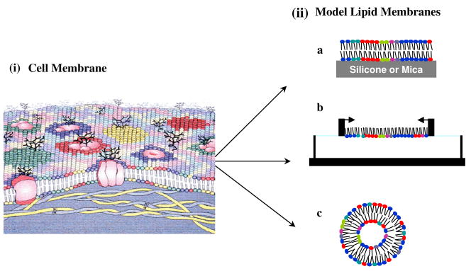
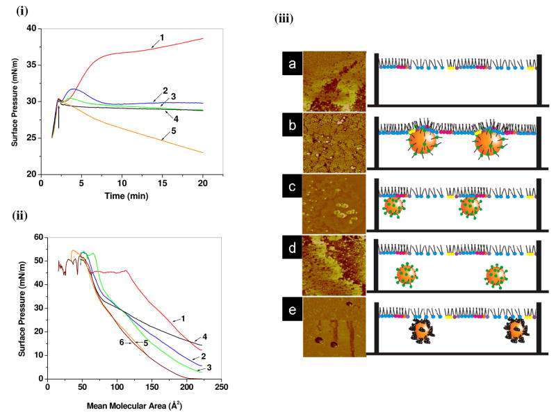
The transport of drugs or drug delivery systems across the cell membrane is a complex biological process, often difficult to understand because of its dynamic nature. In this regard, model lipid membranes, which mimic many aspects of cell-membrane lipids, have been very useful in helping investigators to discern the roles of lipids in cellular interactions. One can use drug-lipid interactions to predict pharmacokinetic properties of drugs, such as their transport, biodistribution, accumulation, and hence efficacy. These interactions can also be used to study the mechanisms of transport, based on the structure and hydrophilicity/hydrophobicity of drug molecules. In recent years, model lipid membranes have also been explored to understand their mechanisms of interactions with peptides, polymers, and nanocarriers. These interaction studies can be used to design and develop efficient drug delivery systems. Changes in the lipid composition of cells and tissue in certain disease conditions may alter biophysical interactions, which could be explored to develop target-specific drugs and drug delivery systems. In this review, we discuss different model membranes, drug-lipid interactions and their significance, studies of model membrane interactions with nanocarriers, and how biophysical interaction studies with lipid model membranes could play an important role in drug discovery and drug delivery.


Figures

Fig. 1
Fig. 1. Schematic illustration of cell membrane and different model membrane systems
(i) This depiction of a cell membrane shows membrane lipid asymmetry as well as microdomains enriched in particular lipids and those induced by membrane proteins. (ii) Model membrane systems mimic the arrangement of lipid in a cell membrane. a, supported lipid bilayer; b, lipid monolayer; c, liposome. Figure 1. (i) Reproduced from ref (6) Copyright 2008 The Authors. Journal compilation copyright 2008 Foundation for Cellular and Molecular Medicine/Blackwell Publishing Ltd.
Fig. 2
Fig. 2. Effect of physiochemical properties of drug on biophysical interactions between drug and model membrane
(i) SP-mean molecular area isotherms of DOPC/DPPC in the presence of ciprofloxacin (A) and moxifloxacin (B), on a subphase of 10 mM Tris at pH 7.4 and 25 °C. Mean molecular areas were corrected to take into account the percentage of fluoroquinolone remaining at the interface. DOPC/DPPC/drug molar ratios were 1:1:0 (continuous line), 1:1:0.4 (discontinuous line), 1:1:1 (dotted line), and 1:1:2 (dash-dotted line). (ii) Effect of fluoroquinolone on the orientation of the DPPC monolayer as revealed by attenuated total reflection-FTIR dichroic spectra. (Top panel) Polarized attenuated total reflection-FTIR spectra of DPPC (a), DPPC/ciprofloxacin (b), and DPPC/moxifloxacin (c). DPPC was used at 50 mg/ml; the molar ratio of lipid:drug was 1:1. Wagging γw(CH2) bands are indicated by arrows. (Bottom panel) Area evolution of dichroic peak of 1200 cm−1 of DPPC in the presence of fluoroquinolone. Integration area of dichroic γw(CH2) band (integrated between 1206 and 1193 cm−1) was plotted versus the DPPC/drug molar ratio as indicated, in the presence of ciprofloxacin (■) and moxifloxacin (●). Reproduced from ref (38). Copyright 2008 by the Biophysical Society.
Fig. 3
Fig. 3. Effect of chemical structure of lipid on biophysical interactions between drug and model membrane
(i) AFM height images (7.5 × 7.5 μm; z-scale: 10 nm) of a mixed DPPC/DOPC (1:1, mol/mol) bilayer recorded in a Tris/NaCl/azithromycin solution at increasing incubation times. (ii) Qualitative evidence for the destabilization of GUVs by azithromycin. Typical fluctuations of a GUV exposed to 50 μM azithromycin over 30 s. Scale bar = 20 μm. Figure 3. (i) Reproduced from ref (41), copyright 2004 Elsevier. Figure 3. (ii) Reproduced from ref (23), copyright 2007 Elsevier.
Figure 4
Figure 4. Effect of physiochemical properties of a drug delivery system on biophysical interactions with model lipid membrane
(i) Interaction of poly(amidoamine) dendrimers with supported lipid bilayers: AFM study. (a) G7 amine terminated dendrimers cause formation of small holes (diameter 15–40 nm) in intact bilayers, (b) G5 amine terminated dendrimers remove lipid molecules from pre-existing defects in lipid bilayers, (c) G5 acetamide dendrimers do not cause hole formation or removal of lipid molecules, but rather adsorb on the edges of existing bilayer defects. (ii) Schematic cross section of the proposed dendrimer-lipid vesicle. Number of dendrimer end-groups (black circles) = M. Number of lipid headgroups in contact with dendrimer surface (inner ring of gray circles) = m. Figure 4. (i) Reproduced from ref (62), copyright 2004 American Chemical Society. Figure 4(ii). Reproduced from ref (64), copyright 2004 American Chemical Society.
Figure 5
Figure 5. Effect of peptide conformation on biophysical interactions of peptide with model lipid membrane
AFM images of LB monolayers composed of (i) Pα, (ii) Pβ with DOPC (A), DOPG (B), DPPC (C), and dipalmitoyl phosphatidylglycerol (D) at a molar ratio of x[5] = 0.15. The scan size is 1 mm × 1 mm, and the z scale for A, B, and D is 3 nm, but for C is 5 nm. Reproduced with permission from ref (69), copyright 2004 American Chemical Society.
Figure 6
Figure 6. Effect of molecular structure of cationic surfactants on biophysical interactions of model lipid membrane with surfactant-modified drug delivery systems
(i) Changes in SP of endothelial cell model membrane (EMM) with time due to interaction with different surfactant-modified NPs. (ii) Changes in the SP-area (π-A) isotherm of the EMM lipid mixture in the presence of different surfactant-modified NPs. (iii) Schematic representation of interaction of (a) EMM-water (control), (b) DMAB-, (c) CTAB- (d) DTAB-, and (e) PVA-modified NPs with EMM. DMAB-modified NPs penetrate into the EMM, whereas CTAB-modified NPs interacts with phospholipids liquid condensed domains only through electrostatic interactions. DTAB- and PVA-modified NPs do not interact. Corresponding AFM phase images of LS films clearly shows that DMAB-modified NPs are embedded in between phospholipids, whereas CTAB-modified NPs are attached to the condensed phospholipid domains. Key: 1, DMAB-NPs; 2, CTAB-NPs; 3, DTAB-NPs; 4, PVA-NPs; 5 unmodified-NPs; 6, EMM alone. Reproduced from ref (75), copyright 2009 American Chemical Society

References

    1. Gorter E, Grendel F. On bimolecular layers of lipoids on the chromocytes of the blood. J Exp Med. 1925;41:439–443. - PMC - PubMed
    1. Singer SJ, Nicolson GL. The fluid mosaic model of the structure of cell membranes. Science. 1972;175:720–731. - PubMed
    1. Escriba PV. Membrane-lipid therapy: a new approach in molecular medicine. Trends Mol Med. 2006;12:34–43. - PubMed
    1. Vereb G, Szollosi J, Matko J, Nagy P, Farkas T, Vigh L, Matyus L, Waldmann TA, Damjanovich S. Dynamic, yet structured: The cell membrane three decades after the Singer-Nicolson model. Proc Natl Acad Sci U S A. 2003;100:8053–8058. - PMC - PubMed
    1. van Meer G, Voelker DR, Feigenson GW. Membrane lipids: where they are and how they behave. Nat Rev Mol Cell Biol. 2008;9:112–124. - PMC - PubMed

Publication types