. 2010 Jan;14(1-2):337-50.
doi: 10.1111/j.1582-4934.2009.00776.x. Epub 2009 May 11.

Co-culture with mesenchymal stromal cells increases proliferation and maintenance of haematopoietic progenitor cells

Affiliations

Co-culture with mesenchymal stromal cells increases proliferation and maintenance of haematopoietic progenitor cells

Thomas Walenda et al. J Cell Mol Med. 2010 Jan.

Abstract

Mesenchymal stromal cells (MSC) have been suggested to provide a suitable cellular environment for in vitro expansion of haematopoietic stem and progenitor cells (HPC) from umbilical cord blood. In this study, we have simultaneously analysed the cell division history and immunophenotypic differentiation of HPC by using cell division tracking with carboxyfluorescein diacetate N-succinimidyl ester (CFSE). Co-culture with MSC greatly enhanced proliferation of human HPC, especially of the more primitive CD34(+)CD38(-) fraction. Without co-culture CD34 and CD133 expressions decreased after several cell divisions, whereas CD38 expression was up-regulated after some cell divisions and then diminished in fast proliferating cells. Co-culture with MSC maintained a primitive immunophenotype (CD34(+), CD133(+) and CD38(-)) for more population doublings, whereas up-regulation of differentiation markers (CD13, CD45 and CD56) in HPC was delayed to higher numbers of cell divisions. Especially MSC of early cell passages maintained CD34 expression in HPC over more cell divisions, whereas MSC of higher passages further enhanced their proliferation rate. Inhibition of mitogen-activated protein kinase 1 (MAPK1) impaired proliferation and differentiation of HPC, but not maintenance of long-term culture initiating cells. siRNA knockdown of N-cadherin and VCAM1 in feeder layer cells increased the fraction of slow dividing HPC, whereas knockdown of integrin beta 1 (ITGB1) and CD44 impaired their differentiation. In conclusion, MSC support proliferation as well as self-renewal of HPC with primitive immunophenotype. The use of early passages of MSC and genetic manipulation of proteins involved in HPC-MSC interaction might further enhance cord blood expansion on MSC.


Figures
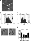
Fig 1
Fig 1
Co-culture with MSC supports proliferation of HPC. Most HPC are elongated upon co-culture with MSC (A). CD34+ cells were stained with CFSE and cultured for 7 days without stromal support (B) or in co-culture with MSC (C, black lines represent fluorescence intensity of non-dividing cells after 24 hrs). The number of cell divisions is indicated for each peak and cells were discerned in a slow dividing fraction (SDF, CFSE+) and a fast dividing fraction (FDF, CFSE). Without stromal support, the fraction of elongated cells was significantly higher in the SDF and this was inversed by co-culture with MSC (D, E; magnification 400×, scale bar 10 μm; *=P < 0.05; ***=P < 0.001; n= 7).
Fig 2
Fig 2
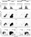
CD34/CD38 expression in relation to the number of HPC divisions. CD34+ cells (left panels) or CD34+CD38 cells (right panels) were stained with CFSE and cultured without or with MSC for 7 days. The number of cell divisions is indicated for each peak (A). CD34 expression is maintained in the CFSE+ cells and decreases after several cell divisions (B). CD38 expression is transiently up-regulated after several cell divisions and decreases thereafter in the FDF (C). Upon co-culture with MSC, the percentage of primitive CD34+CD38 cells was lower, whereas the number of gated events was higher (D).
Fig 3
Fig 3
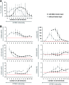
Co-culture shifts differentiation to higher numbers of cell division. CD34+ cells were stained with CFSE and simultaneously cultured either without (grey dots) or with MSC (black dots) for 7 days. Each CFSE-peak (representing 0 to 12 cell divisions) was individually gated and analysed. Co-culture with MSC enhanced the number of cell divisions (A). Decrease of CD34 expression of CD133 and of CD38 expression was delayed to a higher number of cell divisions by co-culture with MSC. On the other hand, up-regulation of differentiation associated markers CD45, CD13 and CD56 shifted to higher numbers of cell division (B). Data of three independent experiments were normalized to the corresponding auto-fluorescence (red lines) and mean ± S.D. are demonstrated.
Fig 4
Fig 4
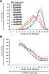
Replicative senescence affects the haematopoiesis supportive function of MSC. MSC of higher passages enhanced proliferation of HPC (blue lines, 9–12 population doublings) in comparison to MSC of early passages (red lines, 5–10 population doublings) (A). Cells remained CD34+ for more cell divisions if cultured on MSC of earlier passage (B).
Fig 5
Fig 5
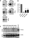
Specific knockdown of adhesion proteins by siRNA. Knockdown of N-cadherin (N-CDH), cadherin-11 (CDH11), integrin beta 1 (ITGB1), CD44 and MAPK1 in MSC was verified after 2 days by Western blot analysis (A). Knockdown of ITGB1, VCAM1 and Jagged1 was validated after 2 days by quantitative RT-PCR (B; **=P < 0.01; ***=P < 0.001). The transient siRNA effect lasted for more than 7 days (C).
Fig 6
Fig 6
The role of various adhesion proteins for stromal function. CD34+ HPC were co-cultured on MSC upon siRNA knockdown of specific proteins. Alternatively, MAPK1 was inhibited by PD098059. The percentage of slow dividing cells (A), CD34+ cells (C) and CD34+CD38 cells (E) was determined after 7 days of co-culture. Means ± S.D. of eight independent experiments are presented in relation to untreated MSC feeder layer. Furthermore, representative results for proliferation (B), CD34 expression (D) and CD38 expression (F) in relation to the number of population doublings (residual CFSE stain) are demonstrated. Despite the increase of CD34+ and CD34+CD38 cells, there was no significant effect on the maintenance of long-term culture-initiating cells (LTC-IC) upon knockdown/inhibition of ITGB1, CD44 or MAPK1 (G; *=P < 0.05; **=P < 0.01; ***=P < 0.001).

Similar articles

Cited by

References

    1. Broxmeyer HE, Douglas GW, Hangoc G, et al. Human umbilical cord blood as a potential source of transplantable hematopoietic stem/progenitor cells. Proc Natl Acad Sci USA. 1989;86:3828–32. - PMC - PubMed
    1. Da Silva CL, Goncalves R, Crapnell KB, et al. A human stromal-based serum-free culture system supports the ex vivo expansion/maintenance of bone marrow and cord blood hematopoietic stem/progenitor cells. Exp Hematol. 2005;33:828–35. - PubMed
    1. Hofmeister CC, Zhang J, Knight KL, et al. Ex vivo expansion of umbilical cord blood stem cells for transplantation: growing knowledge from the hematopoietic niche. Bone Marrow Transplant. 2007;39:11–23. - PubMed
    1. Kohler T, Plettig R, Wetzstein W, et al. Defining optimum conditions for the ex vivo expansion of human umbilical cord blood cells. Influences of progenitor enrichment, interference with feeder layers, early-acting cytokines and agitation of culture vessels. Stem Cells. 1999;17:19–24. - PubMed
    1. Zhang CC, Kaba M, Iizuka S, et al. Angiopoietin-like 5 and IGFBP2 stimulate ex vivo expansion of human cord blood hematopoietic stem cells as assayed by NOD/SCID transplantation. Blood. 2008;111:3415–23. - PMC - PubMed

Publication types

MeSH terms