. 2010 May;14(5):1180-93.
doi: 10.1111/j.1582-4934.2009.00771.x. Epub 2009 May 11.

Lipophilic but not hydrophilic statins selectively induce cell death in gynaecological cancers expressing high levels of HMGCoA reductase

Affiliations

Lipophilic but not hydrophilic statins selectively induce cell death in gynaecological cancers expressing high levels of HMGCoA reductase

S Kato et al. J Cell Mol Med. 2010 May.

Abstract

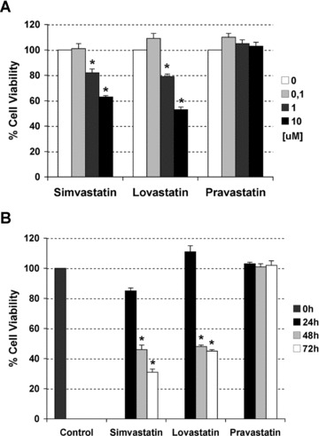
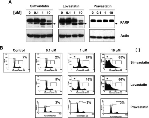
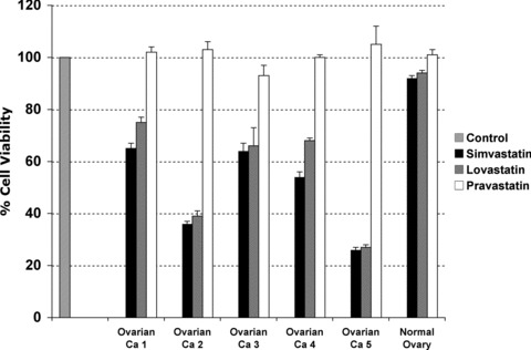
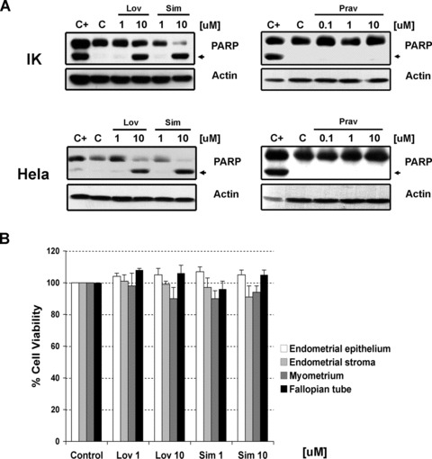
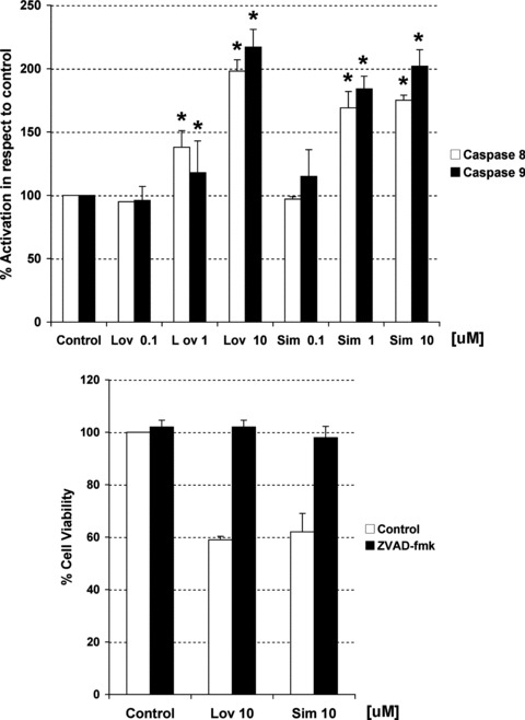
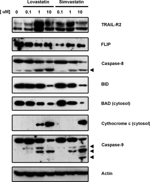
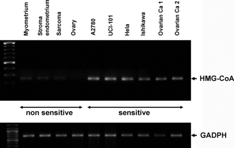
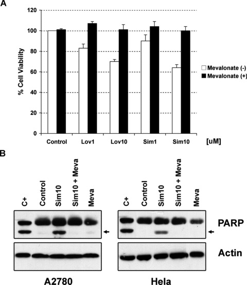
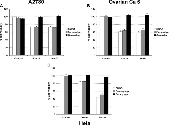
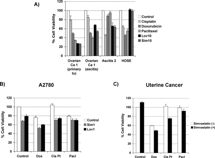
Recent reports have suggested that statins induce cell death in certain epithelial cancers and that patients taking statins to reduce cholesterol levels possess lower cancer incidence. However, little is known about the mechanisms of action of different statins or the effects of these statins in gynaecological malignancies. The apoptotic potential of two lipophilic statins (lovastatin and simvastatin) and one hydrophilic statin (pravastatin) was assessed in cancer cell lines (ovarian, endometrial and cervical) and primary cultured cancerous and normal tissues. Cell viability was studied by MTS assays and apoptosis was confirmed by Western blotting of PARP and flow cytometry. The expressions of key apoptotic cascade proteins were analysed. Our results demonstrate that both lovastatin and simvastatin, but not pravastatin, selectively induced cell death in dose- and time-dependent manner in ovarian, endometrial and cervical cancers. Little or no toxicity was observed with any statin on normal cells. Lipophilic statins induced activation of caspase-8 and -9; BID cleavage, cytochrome C release and PARP cleavage. Statin-sensitive cancers expressed high levels of HMG-CoA reductase compared with resistant cultures. The effect of lipophilic statins was dependent on inhibition of enzymatic activity of HMG-CoA reductase since mevalonate pre-incubation almost completely abrogated the apoptotic effect. Moreover, the apoptotic effect involved the inhibition of synthesis of geranylgeranyl pyrophosphate rather than farnesyl pyrophosphate. In conclusion, lipophilic but not hydrophilic statins induce cell death through activation of extrinsic and intrinsic apoptotic cascades in cancerous cells from the human female genital tract, which express high levels of HMG-CoA reductase. These results promote further investigation in the use of lipophilic statins as anticancer agents in gynaecological malignancies.


Figures

Fig 1
Fig 1
Dose response curve (A) and time course (B) to two lipophilic (lovastatin and simvastatin) and one hydrophilic (pravastatin) statins in A2780 ovarian cancer cells. Cell viability was measured by MTS assays. Data are shown as mean –/+ SD (n= 3). The * indicates statistical significance compared to control (vehicle), (Mann–Whitney test, P-value < 0.05).
Fig 2
Fig 2
(A) Detection by immunoblotting of the cleaved form of PARP, a demonstration that lipophilic (lovastatin and simvastatin) but not hydrophilic (pravastatin) statins induce apoptosis in concentration-dependent manner in A2780 cells. Actin is used as loading control. (B) Determination of sub G0/G1 region en DNA histogram by FACS in A2780 cells treated under same conditions.
Fig 3
Fig 3
Comparative effects in cell survival of lipophilic (lovastatin and simvastatin) and hydrophilic (pravastatin) statins in primary tissue cultures established from ascites of patients with advanced epithelial ovarian carcinoma (indicated as Ovarian Ca 1 to 5) and from normal ovarian epithelium. Cell viability was measured by MTS assays upon 48 hrs of treatment with each statin at 10 uM. Ovarian Ca = Ovarian cancer sample.
Fig 4
Fig 4
(A) Detection by immunoblotting of the cleaved form of PARP after treatment with increasing concentrations of lipophilic (lovastatin and simvastatin) or hydrophilic (pravastatin) statins in Ishikawa (indicated as IK) and Hela cells. Actin is used as loading control. C+ stands for positive control. (B) Absence of effects in cell viability of different concentrations (1–10 uM) of lovastatin (Lov) and simvastatin (Sim) in tissue cultures established from normal gynaecological origin tissues. Cell viability was measured by MTS assays upon 48 hrs of treatment.
Fig 5
Fig 5
(A) Measurement of caspase-8 and -9 activities through an in vitro caspase assay in A2780 cells after 48 hrs of treatment with increasing concentrations of lovastatin (Lov) and simvastatin (Sim). Activity is expressed as per cent from control. (B) Effects in cell viability of both statins in the presence or absence of a non-selective caspase inhibitor (ZVAD-fmk). Data are shown as mean –/+ SD (n= 3). The * indicates statistical significance compared to control (vehicle), (Mann–Whitney test, P-value < 0.05).
Fig 6
Fig 6
Determination by immunoblotting of expression levels of different proteins involved in the extrinsic (TRAIL-R2, FLIP, cleaved caspase-8 and BID) and intrinsic apoptotic cascade (BAD, cytochrome c release and cleaved caspase-9) after incubation with increasing concentrations of lipophilic statins for 24 hrs. Actin was used as loading control.
Fig 7
Fig 7
Determination of basal mRNA levels of HMG-CoA reductase in cell lines and primary tissue cultures from gynaecological origin considered sensitive (ovarian: A2780, UCI-101, ca ovary 1–2; cervical: Hela; endometrial: Ishikawa) and non-sensitive (normal myometrium, endometrial stroma, ovarian tissues and uterine sarcoma) to statin-mediated cell death through RT-PCR. GADPH is shown as loading control.
Fig 8
Fig 8
(A) Effects of increasing concentrations of lipophilic statins in cell viability of A2780 cells in presence or absence of mevalonate (100 uM). (B) Detection by immunoblotting of the cleaved form of PARP in A2780 cells under basal conditions (control) and after treatment with simvastatin (sim10), mevalonate (meva) or their combination for 48 hrs. Actin is shown as loading control. C+ stands for positive control.
Fig 9
Fig 9
(A) Effect of lipophilic (lovastatin: Lov10; simvastatin: Sim10) statins used at 10 uM for 48 hrs in the presence or absence of vehicle (DMSO), geranylgeranyl (geranyl-pp, 10 uM) or farnesyl (farnesyl-pp, 10 uM) pyrophosphates in two cell lines [A2780 (A) and Hela (C)] and one cancerous primary tissue culture [ca ovary 6 (B)]. Cell viability was measured by MTS assay.
Fig 10
Fig 10
(A) Comparative effects in cell survival between treatment with lipophilic statins (lovastatin: lov; simvastatin: sim; used at 10 uM) and chemotherapies (doxorubicin: dox; cisplatin: cis Pt; and paclitaxel: pacl; all used at 5 uM) in three cancerous primary tissue cultures and an immortalized ovarian cell line (HOSE). (B) Effects in cell viability by the combination of statins (lovastatin: lov, and simvastatin: sim, at 1 uM) and different chemotherapies (at 5 uM) in A2780 cells. (C) Similar experiment in a primary tissue culture from recurrent uterine cancer but using lower concentrations of chemotherapies (at 1 uM). Cell viability was measured by MTS assays upon 48 hrs of treatment.

Similar articles

Cited by

References

    1. Heintz AP, Odicino F, Maisonneuve P, et al. Carcinoma of the ovary. FIGO 6th Annual Report on the Results of Treatment in Gynecological Cancer. Int J Gynaecol Obstet. 2006;95:S161–92. - PubMed
    1. Creasman WT, Odicino F, Maisonneuve P, et al. Carcinoma of the corpus uteri. FIGO 6th Annual Report on the Results of Treatment in Gynecological Cancer. Int J Gynaecol Obstet. 2006;95:S105–43. - PubMed
    1. Quinn MA, Benedet JL, Odicino F, et al. Carcinoma of the cervix uteri. FIGO 6th Annual Report on the Results of Treatment in Gynecological Cancer. Int J Gynaecol Obstet. 2006;95:S43–103. - PubMed
    1. Wang CY, Liu PY, Liao JK. Pleiotropic effects of statin therapy: molecular mechanisms and clinical results. Trends Mol Med. 2008;14:37–44. - PMC - PubMed
    1. Beckman JA, Creager MA. The nonlipid effects of statins on endothelial function. Trends Cardiovasc Med. 2006;16:156–62. - PubMed

Publication types

MeSH terms