. 2009 May 11:10:39.
doi: 10.1186/1471-2199-10-39.

Revised Mimivirus major capsid protein sequence reveals intron-containing gene structure and extra domain

Affiliations

Revised Mimivirus major capsid protein sequence reveals intron-containing gene structure and extra domain

Saïd Azza et al. BMC Mol Biol. .

Abstract

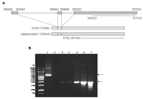
Background: Acanthamoebae polyphaga Mimivirus (APM) is the largest known dsDNA virus. The viral particle has a nearly icosahedral structure with an internal capsid shell surrounded with a dense layer of fibrils. A Capsid protein sequence, D13L, was deduced from the APM L425 coding gene and was shown to be the most abundant protein found within the viral particle. However this protein remained poorly characterised until now. A revised protein sequence deposited in a database suggested an additional N-terminal stretch of 142 amino acids missing from the original deduced sequence. This result led us to investigate the L425 gene structure and the biochemical properties of the complete APM major Capsid protein.

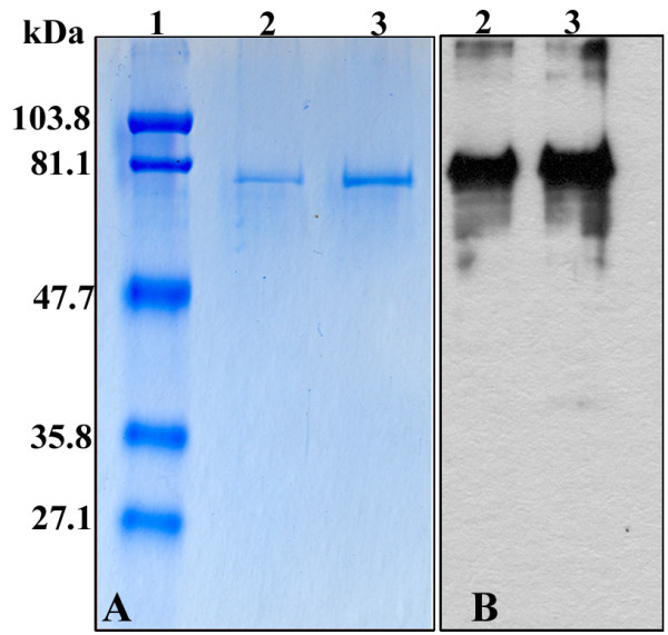
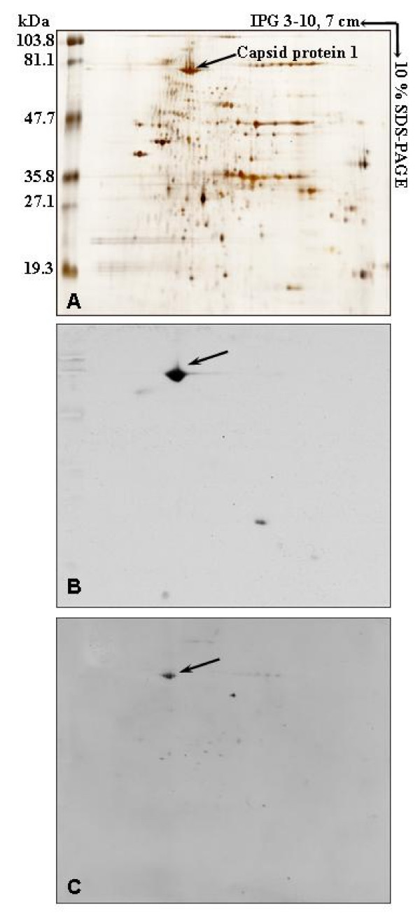
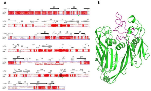
Results: This study describes the full length 3430 bp Capsid coding gene and characterises the 593 amino acids long corresponding Capsid protein 1. The recombinant full length protein allowed the production of a specific monoclonal antibody able to detect the Capsid protein 1 within the viral particle. This protein appeared to be post-translationnally modified by glycosylation and phosphorylation. We proposed a secondary structure prediction of APM Capsid protein 1 compared to the Capsid protein structure of Paramecium Bursaria Chlorella Virus 1, another member of the Nucleo-Cytoplasmic Large DNA virus family.

Conclusion: The characterisation of the full length L425 Capsid coding gene of Acanthamoebae polyphaga Mimivirus provides new insights into the structure of the main Capsid protein. The production of a full length recombinant protein will be useful for further structural studies.


Figures

Figure 1
Figure 1
Schematic organisation and expression of the full length L425 gene. (A) L425 gene (nt 560926 – 557533 position on the APM genomic DNA) showed three coding sequences, boxed in grey, separated by two intron sequences. Organisation of the corresponding cDNA and 593AA full length protein are shown below. Locations of the former D13L capsid coding sequence and corresponding protein are indicated by dotted lines. (B) PCR on APM genomic DNA (lane 1) and RT-PCR analysis on RNA extracted from uninfected (lane 2) or APM-infected amoebae at 0, 2, 4, 8 and 16 h p.i. (lanes 3–7, respectively) using 5' Q5UQL7NcoIF and 3' Q5UQL7SmaIR primers. Left lane: DNA size marker.
Figure 2
Figure 2
Detection of the purified recombinant Capsid protein 1. Either 0.5 or 1 μg of protein (lanes 2 and 3 respectively) was electrophoresed onto 10% SDS-PAGE and revealed by Coomassie blue staining (A) or Western-blot analysis using anti-Histidine mAb. Standard molecular weight markers (lane 1) are indicated on the left.
Figure 3
Figure 3
Two-dimensional gel analysis of APM associated proteins. Viral proteins were revealed by silver staining (A); by Western blot analysis using the Capsid protein 1 specific mAb P9A3 (B). Phosphorylated proteins were revealed with anti-Phosphoserine mAb (C). Standard molecular weight markers are indicated on the left.
Figure 4
Figure 4
Sequence alignment and 3D modelling of APM Capsid protein 1 with that of Chlorella virus PBCV1. (A) Sequence numbering is from the Chlorella virus capsid [PDB:1J5Q]. The putative N-glycosylation sites in APM Capsid are identified by green circles. An insertion of 200 AA replaces a coil motif in 1J5Q (positions 265–315) not belonging to the «jelly-roll» motifs. (B) 3D structure representation of Chlorella virus PBCV1 Capsid core domain (green), and the coil domain (magenta) which is substituted in APM Capsid.

References

    1. La Scola B, Audic S, Robert C, Jungang L, de Lamballerie X, Drancourt M, Birtles R, Claverie JM, Raoult D. A giant virus in amoebae. Science. 2003;299:2033. doi: 10.1126/science.1081867. - DOI - PubMed
    1. Iyer LM, Aravind L, Koonin EV. Common origin of four diverse families of large eukaryotic DNA viruses. J Virol. 2001;75:11720–11734. doi: 10.1128/JVI.75.23.11720-11734.2001. - DOI - PMC - PubMed
    1. Raoult D, Audic S, Robert C, Abergel C, Renesto P, Ogata H, La Scola B, Suzan M, Claverie JM. The 1.2-megabase genome sequence of Mimivirus. Science. 2004;306:1344–1350. doi: 10.1126/science.1101485. - DOI - PubMed
    1. Suzan-Monti M, La Scola B, Barrassi L, Espinosa L, Raoult D. Ultrastructural characterization of the giant volcano-like virus factory of Acanthamoeba polyphaga Mimivirus. PLoS ONE. 2007;2:e328. doi: 10.1371/journal.pone.0000328. - DOI - PMC - PubMed
    1. Xiao C, Chipman PR, Battisti AJ, Bowman VD, Renesto P, Raoult D, Rossmann MG. Cryo-electron microscopy of the giant Mimivirus. J Mol Biol. 2005;353:493–496. doi: 10.1016/j.jmb.2005.08.060. - DOI - PubMed

Publication types