. 2009 May 11:8:1.
doi: 10.1186/1476-5926-8-1.

Low affinity glucocorticoid binding site ligands as potential anti-fibrogenics

Affiliations

Low affinity glucocorticoid binding site ligands as potential anti-fibrogenics

Carylyn J Marek et al. Comp Hepatol. .

Abstract

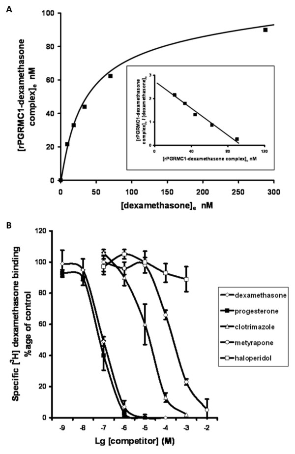
Background: Pregnane X receptor (PXR) agonists inhibit liver fibrosis. However, the rodent PXR activator pregnenolone 16alpha carbonitrile (PCN) blocks, in vitro, hepatic stellate cell-to-myofibroblast trans-differentiation and proliferation in cells from mice with a disrupted PXR gene, suggesting there is an additional anti-fibrogenic drug target for PCN. The role of the low affinity glucocorticoid binding site (LAGS) - which may be identical or associated with the progesterone receptor membrane component 1 (PGRMC1) - in mediating this anti-fibrogenic effect has been examined, since binding of dexamethasone to the LAGS in liver microsomal membranes has previously been shown to be inhibited by PCN.

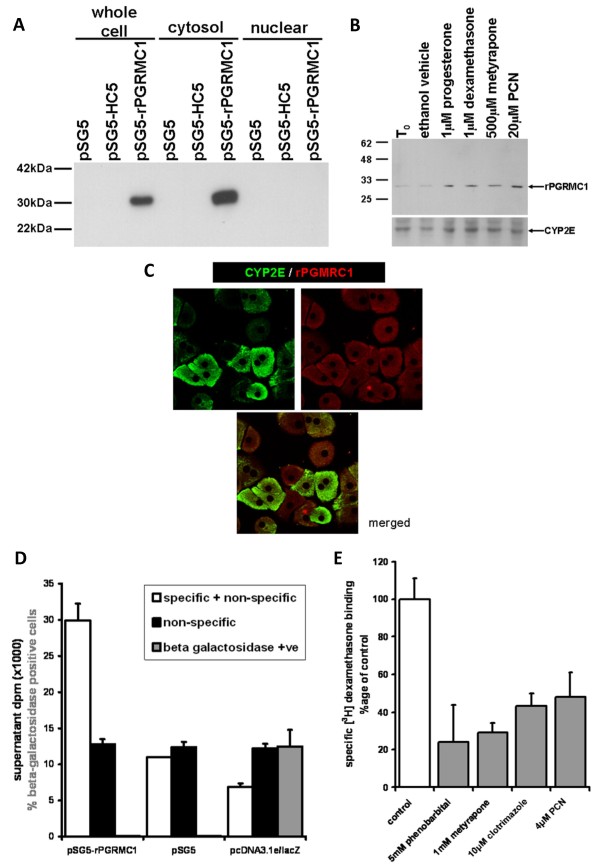
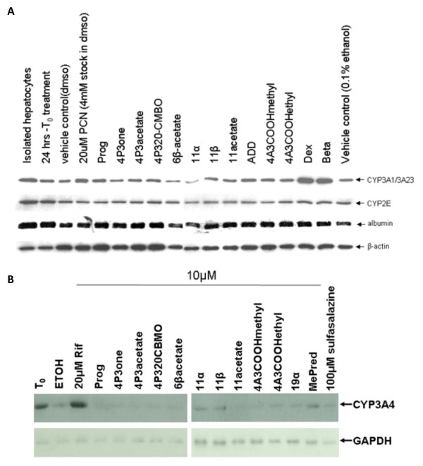
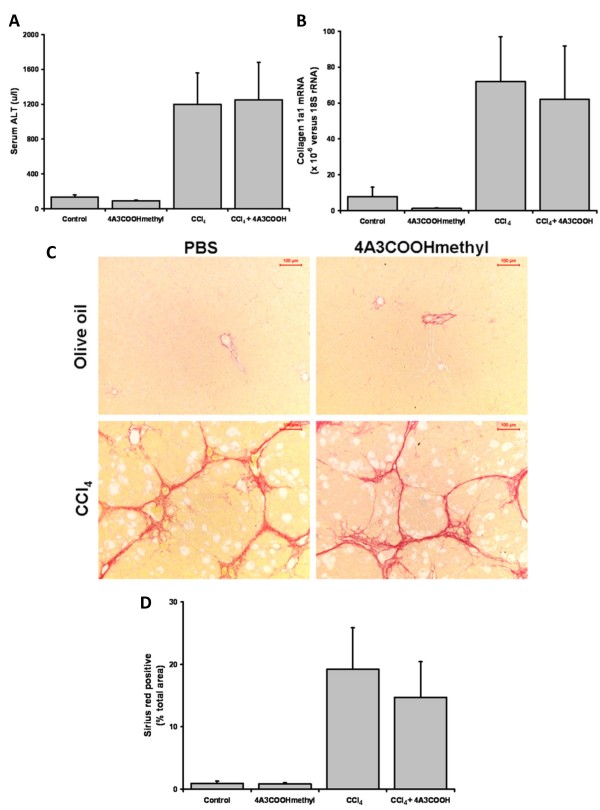
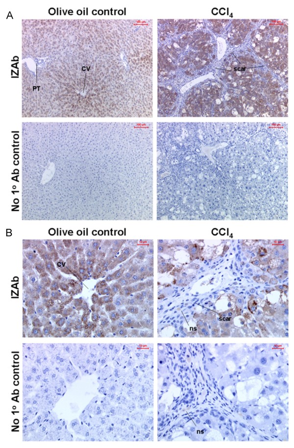
Results: Quiescent rat and human hepatic stellate cells (HSC) were isolated from livers and cultured to generate liver myofibroblasts. HSC and myofibroblasts expressed PGRMC1 as determined by RT-PCR and Western blotting. Quiescent rat HSC also expressed the truncated HC5 variant of rPGRMC1. Rat PGRMC1 was cloned and expression in COS-7 cells gave rise to specific binding of radiolabelled dexamethasone in cell extracts that was inhibited by PCN, suggesting that PGRMC1 may be identical to LAGS or activates LAGS binding activity. Liver microsomes were used to screen a range of structurally related compounds for their ability to inhibit radiolabelled dexamethasone binding to rat LAGS. These compounds were also screened for their ability to activate rat and human PXR and to inhibit rat HSC-to-myofibroblast trans-differentiation/proliferation. A compound (4 androstene-3-one 17beta-carboxylic acid methyl ester) was identified which bound rat LAGS with high affinity and inhibited both rat and human HSC trans-differentiation/proliferation to fibrogenic myofibroblasts without showing evidence of rat or human PXR agonism. However, despite potent anti-fibrogenic effects in vitro, this compound did not modulate liver fibrosis severity in a rat model of liver fibrosis. Immunohistochemical analysis showed that rat liver myofibroblasts in vivo did not express rPGRMC1.

Conclusion: LAGS ligands inhibit HSC trans-differentiation and proliferation in vitro but show little efficacy in inhibiting liver fibrosis, in vivo. The reason(s) for this disparity is/are likely associated with an altered myofibroblast phenotype, in vitro, with expression of rPGMRC1 in vitro but not in vivo. These data emphasize the limitations of in vitro-derived myofibroblasts for predicting their activity in vivo, in studies of fibrogenesis. The data also demonstrate that the anti-fibrogenic effects of PCN in vivo are likely mediated entirely via the PXR.


Figures

Figure 1
Figure 1
Rat and human HSCs and myofibroblasts express PGRMC1 in vitro. Left panel, RT-PCR analysis for rPGRMC1 in rat cells and tissues as indicated using primer sequences and conditions as outlined in methods section. T6 cells are a rat hepatic stellate cell line [47] (a). Right panel, Western blot of the indicated cell types for rPGRMC1 using the anti-IZAb (a). RT-PCR products from the indicated cell types with and without digestion with the restriction enzyme Nci-I as indicated. rPGRMC1 PCR product does not contain an Nci-I site whereas the truncated HC5 variant contains a single site and is cleaved [17] (b). Left panel, RT-PCR analysis for hPGRMC1 in human cells using primer sequences and conditions as outlined in methods section. Senescent myofibroblasts had ceased proliferation (typically at passage 3–5) (c). Right panel, Western blot of the indicated anonymised donor cells for hPGRMC1 using the anti-IZAb (c). Results typical of a least 3 independent experiments and/or animals except right panel (c), 2 separate human donors.
Figure 2
Figure 2
Expression of rPGRMC1 results in dexamethasone binding activity. Western blot for rPGRMC1 in various cell fractions using the anti-IZ Ab in COS-7 cells transfected with the indicated construct. All lanes were loaded with 10 μg protein/lane. Note, HC5 is a truncated form of rPGRMC1 cloned from rat kidney [17] (a). Western blot for rPGRMC1 using the anti-IZ Ab and CYP2E1 (lower blot). Rat hepatocytes were cultured for 24 hours to allow attachment (T0) and then treated for 24 hours with the indicated ligand or ethanol vehicle prior to analysis. Each lane contains 10 μg total protein/well, typical of 3 separate experiments (b). Confocal microscopy of rat hepatocytes demonstrating non-nuclear location of PGRMC1 and CYP2E1 (c). 200 × 106 COS-7 cells were transfected with pSG5-rPGRMC1, pSG5 or pcDNA3.1e/lacZ and 13,000 g cell extracts prepared and incubated with radiolabelled dexamethasone as outlined in methods section. Supernatant dpm after charcoal dextran treatment to remove free radioligand is given in dpm after normalisation of protein for total (specific and non-specific) – white bars; and non specific (by co-incubation of 1000-fold molar excess unlabelled dexamethasone) – black bars. The percentage of cells that stained positive for beta galactosidase activity (grey bars) was determined in situ in separate wells by examining at least 5 randomly selected low power fields. Data are the mean and standard deviation of at least 3 separate determinations from the same experiment, typical of 2 separate experiments (d). 200 × 106 COS-7 cells were transfected with pSG5-rPGRMC1. Dexamethasone binding activity was determined in whole COS-7 cells as outlined in methods section and in the presence of the indicated concentration of unlabelled potential competitor. Specific binding was determined by co-incubation of replicates also containing unlabelled 1000-fold molar excess of unlabelled dexamethasone. Typically, non specific binding accounted for between 40–60% of total binding of radioligand. Data are the mean and standard deviation of 3 separate determinations from the same experiment, typical of 3 separate experiments. Control is the mean and standard deviation specific activity of 3 determinations from the same experiment after subtraction of non-specific binding. The percent of binding in the presence of unlabelled competitors was determined after subtraction of non-specific binding. Data are typical of at least 2 separate experiments (e).
Figure 3
Figure 3
Radiolabelled dexamethasone interacts in a specific and saturable manner with rat liver microsomes and binding is competed by selected compounds. Male rat liver microsomes were incubated in duplicate with increasing concentrations of radiolabelled dexamethasone (ligand) with or without excess unlabelled dexamethasone and allowed to reach equilibrium on ice. A small volume of each incubation was removed to determine the total ligand concentration ([L0]) prior to removal of free unbound ligand by dextran/charcoal adsorption. Specifically bound ligand at equilibrium ([LRe]) was calculated by subtracting radioactive counts present in samples which also contained excess unlabelled dexamethasone after dextran-charcoal adsorption (and was typically < 5%). Free ligand concentration at equilibrium was calculated by subtracting specifically bound ligand at equilibrium from the total ligand concentration (i.e. [L0] - [LRe]) and assumes receptor-ligand stoichiometry of 1:1. Results typical of six separate preparations (a). Male rat liver microsomes were incubated with 50 nM [3H] dexamethasone as outlined in methods section with or without excess unlabelled dexamethasone (to determine non-specific binding) or a range of unlabelled compounds (added with ethanol vehicle such that final ethanol concentration was 1%, also present in controls). After overnight incubation on ice, free ligand was removed by dextran-charcoal adsorption and specifically bound radiolabelled dexamethasone determined (b).
Figure 4
Figure 4
Screening for PXR activators in rat and human hepatocytes via CYP3A induction. Rat hepatocytes were isolated and cultured as outlined in methods section. After 24 hours of culture (T0), hepatocytes were treated for a further 24 hours with 10 μM of the indicated compound from a 1000 fold ethanol-solvated stock (except PCN, which was added to give 20 μM from a DMSO-solvated stock). Equivalent ethanol (0.1% v/v) and DMSO (0.5% v/v) vehicles are included. Cells were then analyzed for expression of the indicated protein by Western blotting, 10 μg total protein/lane. Results are typical of at least 3 separate experiments (a). Human hepatocytes were treated essentially as for rat hepatocytes except that all compounds were prepared as ethanol solvated stocks. Cells were then analyzed for expression of the indicated protein by Western blotting, 20 μg total protein/lane. Results are from one donor (LH2), typical of 2 different donors (b).
Figure 5
Figure 5
Screening for inhibitors of trans-differentiation in rat and human HSCs – Part 1. Rat HSCs were isolated and cultured for 2 days (T0) whereupon cells were treated with the indicated compound as outlined in methods section. After 9 days, cells were analyzed by Western blotting for α-smooth muscle actin (α-sma). Each lane contains 10 μg total protein/lane, results typical of at least 3 separate experiments (a). Human HSCs were treated with the indicated compound and confluence determined in randomly selected fields. Data are the mean and standard deviation confluence at day 12 of 3 separate treatment dishes from the same donor, typical of at least 3 separate donors (b).
Figure 6
Figure 6
Screening for inhibitors of trans-differentiation in rat and human HSCs – Part 2. Photomicrographs of human HSCs at day 12 of culture with the indicated treatment (a); levels of expression of α-smooth muscle actin (α-sma) (b) and levels of expression of fibrogenic marker mRNA transcripts in human HSCs after treatment selected compounds for 19 days. Data are the mean and standard deviation relative transcript level from 3 separate treatments on cells from the same donor, typical of at least 3 separate donors (c).
Figure 7
Figure 7
4A3COOHmethyl administration and liver fibrosis in a rat CCl4 model of liver fibrosis. Four animals/group (control or 4A3COOHmethyl) or six animals/group (CCl4 or CCl4 + 4A3COOHmethyl) were treated as outlined in the Methods section. Mean and standard deviation serum ALT (a); Mean and standard deviation collagen 1A1 mRNA levels (b); typical views of liver sections stained for sirius red, with a 100 μm scale bar (b); quantitative image analysis for fibrosis – data are the mean and standard deviation percentage sirius red staining from at least 4 separate animals in each treatment with at least 10 randomly selected fields examined for each animal (c).
Figure 8
Figure 8
Rat liver myofibroblast do not express rPGRMC1 in vivo – Part A. Low power views (a) and high power views (b) of liver section immunohistochemically stained for rPGRMC1 using IZAb upper panels or identical staining without addition of IZAb (no 1° Ab control) from olive oil control or CCl4 treated animals (note CCl4 + 4A3COOHmethyl treated animals gave similar results). PT, portal tract; CV, central vein; scar, primary location of scar matrix and liver myofibroblasts; ns non-specifically bound secondary antibody.
Figure 9
Figure 9
Rat liver myofibroblast do not express rPGRMC1 in vivo – Part B. High powered views show positive staining of non-parenchymal cells in control liver sections (a); co-staining sections from indicated treatment groups – DNA with DAPI (blue), α-sma (green) and PGRMC1 with IZAb (red) with merged panel (b); high powered view of merged liver section from CCl4-treated rat liver (c). PV, periportal venule; PA, periportal arteriole; BD, bile duct.

Similar articles

Cited by

References

    1. Wallace K, Burt AD, Wright MC. Liver fibrosis. Biochem J. 2008;411:1–18. doi: 10.1042/BJ20071570. - DOI - PubMed
    1. Fallowfield JA, Mizuno M, Kendall TJ, Constandinou CM, Benyon RC, Duffield JS, Iredale JP. Scar-associated macrophages are a major source of hepatic matrix metalloproteinase-13 and facilitate the resolution of murine hepatic fibrosis. J Immunol. 2007;178:5288–5295. - PubMed
    1. Kliewer SA, Moore JT, Wade L, Staudinger JL, Watson MA, Jones SA, McKee DD, Oliver BB, Willson TM, Zetterström RH, Perlmann T, Lehmann JM. An orphan nuclear receptor activated by pregnanes defines a novel steroid signaling pathway. Cell. 1998;92:73–82. doi: 10.1016/S0092-8674(00)80900-9. - DOI - PubMed
    1. Goodwin B, Gauthier KC, Umetani M, Watson MA, Lochansky MI, Collins L, Leitersdorf E, Mangelsdorf DJ, Kliewer SA, Repa JJ. Identification of bile acid precursors as endogenous ligands for the nuclear xenobiotic pregnane X receptor. Proc Natl Acad Sci USA. 2003;100:223–228. doi: 10.1073/pnas.0237082100. - DOI - PMC - PubMed
    1. Goodwin B, Redinbo MR, Kliewer SA. Regulation of cyp3a gene transcription by the pregnane x receptor. Annu Rev Pharmacol Toxicol. 2002;42:1–23. doi: 10.1146/annurev.pharmtox.42.111901.111051. - DOI - PubMed

LinkOut - more resources