

Review
doi: 10.1016/j.febslet.2009.05.005.
Epub 2009 May 9.
Glimpses of the molecular mechanisms of beta2-microglobulin fibril formation in vitro: aggregation on a complex energy landscape
Affiliations
- PMID: 19433089
- PMCID: PMC2734061
- DOI: 10.1016/j.febslet.2009.05.005
Item in Clipboard
Review
Glimpses of the molecular mechanisms of beta2-microglobulin fibril formation in vitro: aggregation on a complex energy landscape
FEBS Lett.
.
Abstract
Beta(2)-microglobulin (beta(2)m) is a 99-residue protein that aggregates to form amyloid fibrils in dialysis-related amyloidosis. The protein provides a powerful model for exploration of the structural molecular mechanisms of fibril formation from a full-length protein in vitro. Fibrils have been assembled from beta(2)m under both low pH conditions, where the precursor is disordered, and at neutral pH where the protein is initially natively folded. Here we discuss the roles of sequence and structure in amyloid formation, the current understanding of the structural mechanisms of the early stages of aggregation of beta(2)m at both low and neutral pH, and the common and distinct features of these assembly pathways.
Figures
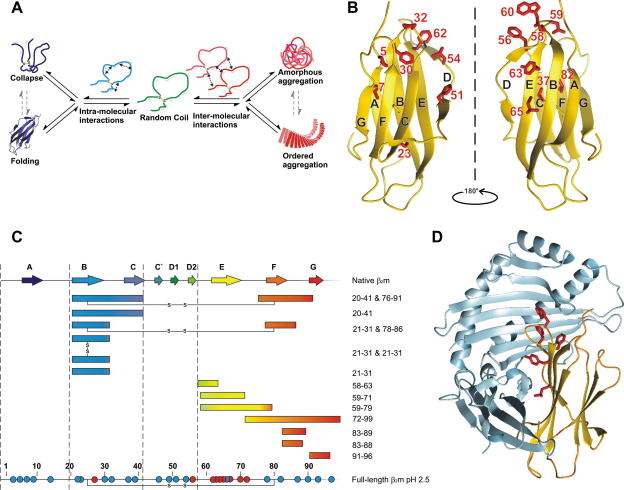
(A) Schematic illustration of the competition between intra-molecular and inter-molecular interactions in protein folding and assembly. When intra-molecular interactions prevail, proteins fold or form collapsed states (left side). By contrast, when inter-molecular interactions dominate, protein aggregation results (right side). (B) Monomeric β2m with residues discussed herein highlighted. (C) Amyloid forming properties of the sequence of β2m when isolated as peptides [10–15], or in the context of the full-length protein (the latter at pH 2.5). Red circles indicate residues that affect fibril formation kinetics, blue circles indicate positions where changes had little or no effect on kinetics . (D) Structure of β2m (gold) in the MHC-1 complex. Hydrophobic residues present in the β-strand E region of β2m (Phe56, Trp60, Tyr62, Tyr63 and Leu65) that contact the heavy chain of the MHC-1 are highlighted.
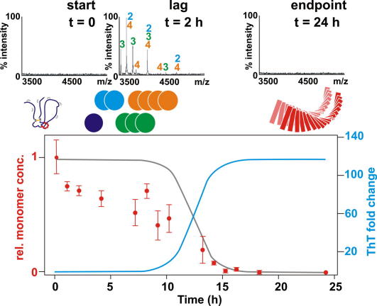
Species formed en route to amyloid fibrils by β2m at pH 2.5 as monitored by mass spectrometry and ThT fluorescence. During the lag phase (blue line, bottom figure), the concentration of monomeric protein (red circles) decreases more rapidly than expected based on ThT data (grey line). ESI-MS data (top) shows the presence of dimers, trimers and tetramers as well as monomers, but no larger species, during the lag phase. At the conclusion of the reaction no oligomers remain. Data were taken from Smith et al. .
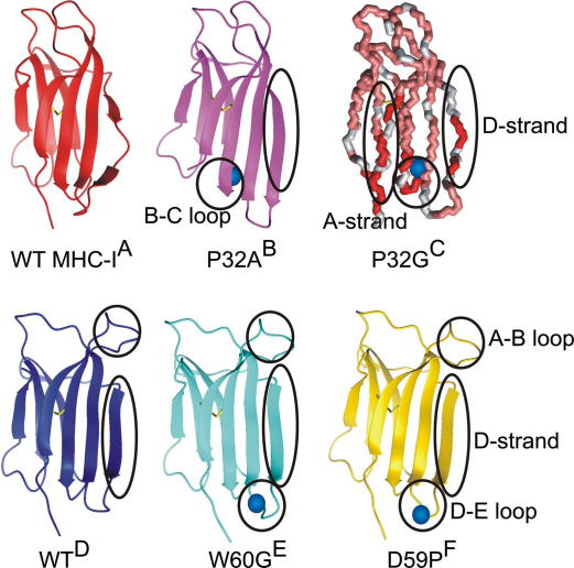
Structures of wild-type β2m and several variants mentioned herein, blue spheres show the site of mutation where relevant. Regions of major conformational change are highlighted. (A) WT β2m in the MHC-1 complex (PDB: 1DUZ) . (B) P32A variant of β2m that crystallises as a dimer and forms digomers in the presence of Cu2+ (2F8O) . (C) Model of structural changes in a folding intermediate of β2m, populated at high levels in the P32G variant, mapped onto the NMR structure of wild-type β2m. Regions that are most perturbed in comparison with wild-type β2m are shown in red, those that show little perturbation are pink and no information is available for those in grey . (D) X-ray crystallographic structure of a rare monomeric species of wild-type β2m with a straight β-strand D (1LDS) . (E) W60G variant of β2m that has a decreased amyloid propensity (2Z9T) . (F) D59P β2m displays an increased propensity to form amyloid (3DHJ) .
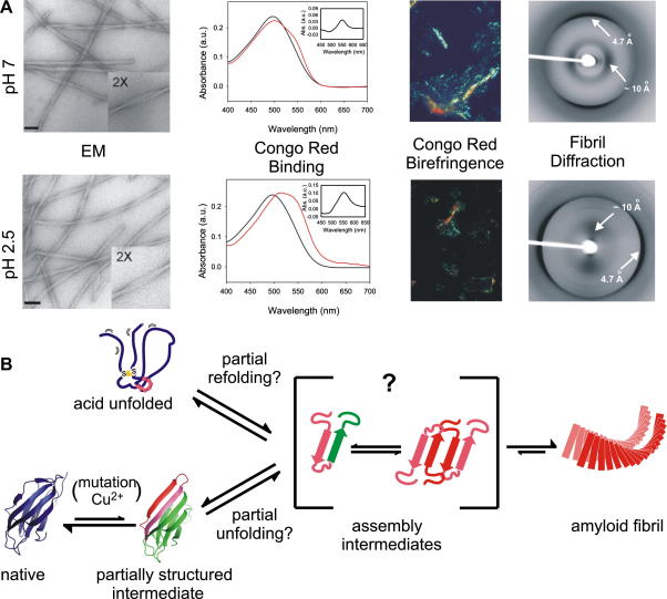
(A) Comparison of experimental data for fibrils of β2m formed at pH 7.0 and 2.5. EM scale bar indicates 100 nm. Absorbance spectra of Congo Red free in solution (black line) and bound to β2m fibrils (red line) as well as the difference spectra (inset). Both fibril types give rise to red-green birefringence in the presence of Congo Red. The X-ray fibre diffraction patterns show reflections at 4.7 Å and ∼10 Å, consistent with a cross-β structure. Data were taken from Jahn . (B) Scheme for convergence of the mechanisms of fibril formation at pH 2.5 and 7.0. Regions with high amyloidogenic propensity are displayed in pink. It is not known precisely how oligomers stack or whether they have ordered β-sheet, however, increased intermolecular protein–protein interactions (red) may be important in the reaction pathway.
References
-
- Sipe J.D., Cohen A.S. Review: history of the amyloid fibril. J. Struct. Biol. 2000;130:88–98. - PubMed
-
- Tartaglia G.G., Pawar A.P., Campioni S., Dobson C.M., Chiti F., Vendruscolo M. Prediction of aggregation-prone regions in structured proteins. J. Mol. Biol. 2008;380:425–436. - PubMed
-
- Westermark P. A primer of amyloid nomenclature. Amyloid. 2007;14:179–183. - PubMed
-
- Dobson C.M. Protein folding and misfolding. Nature. 2003;426:884–890. - PubMed
-
- Saper M.A., Bjorkman P.J., Wiley D.C. Refined structure of the human histocompatibility antigen HLA-A2 at 2.6 Å resolution. J. Mol. Biol. 1991;219:277–319. - PubMed
Publication types
MeSH terms
Substances
Grants and funding
LinkOut - more resources
Full Text Sources
Research Materials
