. 2009 May 18;185(4):743-54.
doi: 10.1083/jcb.200901129. Epub 2009 May 11.

Decorin is a novel antagonistic ligand of the Met receptor

Affiliations

Decorin is a novel antagonistic ligand of the Met receptor

Silvia Goldoni et al. J Cell Biol. .

Abstract

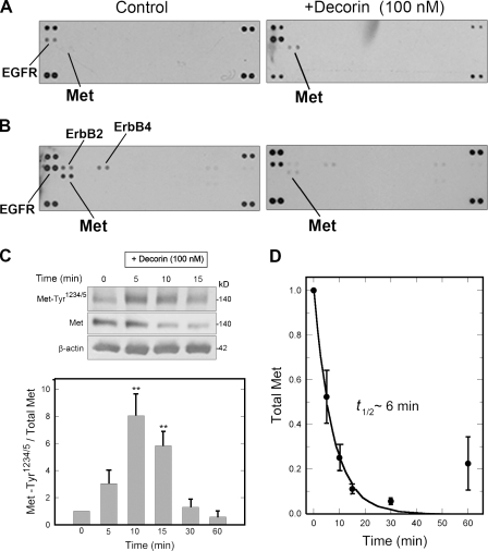
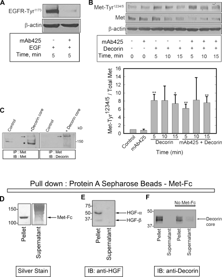
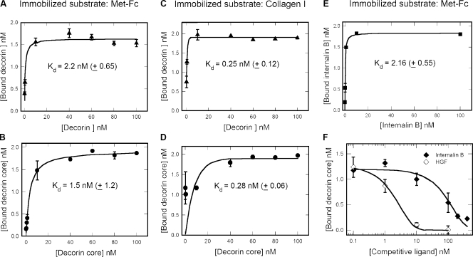
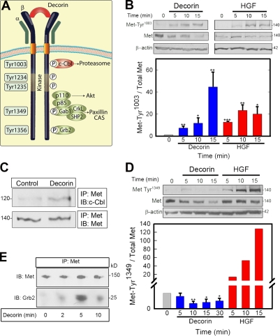
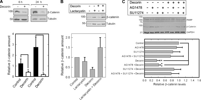
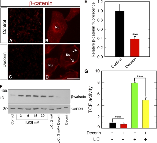
Decorin, a member of the small leucine-rich proteoglycan gene family, impedes tumor cell growth by down-regulating the epidermal growth factor receptor. Decorin has a complex binding repertoire, thus, we predicted that decorin would modulate the bioactivity of other tyrosine kinase receptors. We discovered that decorin binds directly and with high affinity (K(d) = approximately 1.5 nM) to Met, the receptor for hepatocyte growth factor (HGF). Binding of decorin to Met is efficiently displaced by HGF and less efficiently by internalin B, a bacterial Met ligand. Interaction of decorin with Met induces transient receptor activation, recruitment of the E3 ubiquitin ligase c-Cbl, and rapid intracellular degradation of Met (half-life = approximately 6 min). Decorin suppresses intracellular levels of beta-catenin, a known downstream Met effector, and inhibits Met-mediated cell migration and growth. Thus, by antagonistically targeting multiple tyrosine kinase receptors, decorin contributes to reduction in primary tumor growth and metastastic spreading.


Figures

Figure 1.
Figure 1.
Decorin affects Met receptor signaling and turnover. (A) Phospho-RTK arrays. HeLa cells were treated with decorin for 15 min. RTK membranes were incubated with cell lysates. The duplicate dots at each corner represent phospho-Tyr positive controls. (B) The same experiment as in A using nonquiescent cells. (C, top) Representative immunoblot of a short decorin time course showing phosphorylation of the Met receptor at Tyr1234/5, total Met, and β-actin. (bottom) Quantification of immunoblots similar to those shown in the top from three independent experiments performed in triplicate. Values represent the mean ± SEM (**, P < 0.01). (D) Best-fit plot of Met receptor degradation over time. Relative values were obtained by scanning densitometry (chemiluminescence) of blots as in C and represent means ± SEM from three independent experiments performed in triplicate.
Figure 2.
Figure 2.
Decorin interacts with the Met receptor. (A) 1 µg/ml mAb425, an EGFR-specific blocking antibody, was tested before experiments in combination with decorin by evaluating its effect in inhibiting EGF-dependent (16 nM) EGFR phosphorylation. (B, top) Immunoblot of a short decorin (100 nM) time course showing phosphorylation of the Met receptor at Tyr1234/5, total Met, and β-actin in the presence or absence of 1 µg/ml mAb425. (bottom) Quantification of immunoblots similar to those shown in the top panel. Values represent the mean ± SEM from three independent experiments performed in triplicate (*, P < 0.05; **, P < 0.01). (C) Immunoblots detecting Met (left) and decorin (right) in cells treated with decorin protein core for 15 min, cross-linked with 500 nM S-SMPB for 20 min at 37°C, and immunoprecipitated with an anti–C terminus Met antibody. Arrows point to a high Mr complex of Met and decorin protein core (∼190 kD). The asterisk indicates Met monomer (∼140 kD). (D) Silver-stained gel. Notice that the entire Met-Fc is bound to the protein A–Sepharose beads. Smear is the carrier proteins. (E) Immunoblotting (IB) of HGF after pull-down with protein A beads–Met-Fc. (F) Immunoblotting of decorin after pull-down with either protein A beads–Met-Fc or beads alone. Note the absence of HGF or decorin in the supernatants, indicating that essentially all of the ligands were bound. IP, immunoprecipitation. (D–F) Values shown are given in kiloDaltons.
Figure 3.
Figure 3.
Affinity interaction between decorin and the Met receptor. (A–E) Ligand-binding assays using decorin, decorin protein core, or internalin B as soluble ligands and Met-Fc or fibrillar collagen I as immobilized substrates. (F) Competition experiments using constant amounts of decorin core (10 nM) and increasing amounts of internalin B or HGF as indicated. Notice that only at high molar ratios (20:1 and 40:1), internalin B significantly (∼70%) reduces decorin protein core binding to the Met (IC50 = ∼180 nM). In contrast, HGF is much more efficient (IC50 = ∼2.5 nM) in displacing decorin core. Values represent the mean ± SEM.
Figure 4.
Figure 4.
Decorin induces differential and selective phosphorylation of Met Tyr residues. (A) Diagram of the main Met receptor Tyr phosphorylation sites and adaptor proteins. CAS, Crk-associated substrate; P, phosphate. (B, top) Representative immunoblots of a short decorin (100 nM) time course showing phosphorylation of the Met receptor at Tyr1003 and total Met amount vis à vis 1.5 nM HGF. (bottom) Quantification of immunoblots from three independent experiments. (C) Coimmunoprecipitation of c-Cbl and Met using an antibody directed toward the intracellular domain of Met. 100 nM decorin treatment was performed for 10 min. (D, top) Representative immunoblot showing phosphorylation of the Met receptor at Tyr1349 and total Met after 100 nM decorin treatment vis à vis 1.5 nM HGF. (bottom) Quantification of immunoblots from three independent experiments performed in triplicate. (E) Recruitment of Grb2 to the Met receptor mediated by 100 nM decorin. Coimmunoprecipitation of the Met receptor and Grb2 using an anti-Met C terminus antibody for the immunoprecipitation (IP) and either the same antibody or an anti-Grb2 monoclonal antibody for the immunoblotting (IB). Values represent the mean ± SEM (*, P < 0.05; **, P < 0.01; ***, P < 0.001). All of the relative values were obtained by scanning densitometry (chemiluminescence). (B–E) Values shown are given in kiloDaltons.
Figure 5.
Figure 5.
Decorin down-regulates β-catenin via the Met receptor. (A, top) Representative immunoblots of HeLa cells treated with 100 nM decorin for the times indicated and probed for β-catenin. Tubulin was used as a loading control. (bottom) Quantification of immunoblots as those presented in the top panel from three independent experiments. (B, top) β-Catenin levels after treatment with 100 nM decorin for 30 min in the presence or absence of 10 µM of the proteasome inhibitor lactacystin. Cells were preincubated with lactacystin for 1 h before adding decorin. (bottom) Quantification of immunoblots as those presented in the top panel from three independent experiments. (C, top) β-Catenin and PARP immunoblots after treatment with 100 nM decorin for 6 h in the presence or absence of the tyrosine kinase inhibitors AG1478 and SU11274 (both 1 µM) as indicated. Glyceraldehyde-3-phosphate dehydrogenase (GAPDH) was used as loading control. (bottom) Quantification of immunoblots as those presented in the top panel from three independent experiments performed in triplicate. Values represent the mean ± SEM (*, P < 0.05; **, P < 0.01). All of the relative values were obtained by scanning densitometry (chemiluminescence). Values shown in blots are given in kiloDaltons.
Figure 6.
Figure 6.
Decorin attenuates β-catenin levels and transcriptional activity. (A–D) Representative β-catenin immunofluorescence images of HeLa cells after a 2-h incubation with or without 100 nM decorin. Notice the marked decline in β-catenin levels throughout the cytoplasm and perinuclear regions in the decorin-treated cells. In contrast, the plasma membrane localization of β-catenin increases (D, arrows). Nu, nucleus. Bars: (A and C) 50 µm; (B and D) 10 µm. (E) Quantification of the fluorescence intensity of images similar to those shown in A and C. The values represent the mean ± SEM of 12 images (∼120 cells/image) from four independent experiments. (F) Decorin inhibits β-catenin activity via a GSK3-β–independent mechanism. Representative β-catenin immunoblot of HeLa lysates treated with 100 nM decorin for 6 h in the presence or absence of 30 mM of the GSK3-β inhibitor LiCl. Cells were preincubated with LiCl for 1 h in full serum before decorin treatment. Glyceraldehyde-3-phosphate dehydrogenase (GAPDH) was used as a loading control. (G) HeLa cells were cotransfected with the TopFlash vector and a vector carrying the R. reniformis luciferase. 12 h after transfection, cells were treated with or without 100 nM decorin or LiCl for 6 h. Luciferase activity was measured after incubation with the cognate substrate luciferin. The values were normalized on R. reniformis luciferase activity. Cells preincubated with 30 mM LiCl, as in F, showed the same degree of reduction in luciferase activity when treated with decorin. Notice that LiCl enhances β-catenin activity (***, P < 0.001). The values represent the mean ± SEM of five independent experiments performed in triplicate. TCF, T cell factor.

References

    1. Aberle H., Bauer A., Stappert J., Kispert A., Kemler R. 1997. β-catenin is a target for the ubiquitin-proteasome pathway.EMBO J. 16:3797–3804 - PMC - PubMed
    1. Athauda G., Giubellino A., Coleman J.A., Horak C., Steeg P.S., Lee M.-J., Trepel J., Wimberly J., Sun J., Coxon A., et al. 2006. c-Met ectodomain shedding rate correlates with malignant potential.Clin. Cancer Res. 12:4154–4162 - PubMed
    1. Basilico C., Arnesano A., Galuzzo M., Comoglio P.M., Michieli P. 2008. A high affinity hepatocyte growth factor-binding site in the immunoglobulin-like region of Met.J. Biol. Chem. 283:21267–21277 - PMC - PubMed
    1. Berthou S., Aebersold D.M., Schimdt L.S., Stroka D., Heigl C., Streit B., Stalder D., Gruber G., Liang S., Howlett A.R., et al. 2004. The Met kinase inhibitor SU11274 exhibits a selective inhibition pattern toward different receptor mutated variants.Oncogene. 23:5387–5393 - PubMed
    1. Bi X., Tong C., Dokendorff A., Banroft L., Gallagher L., Guzman-Hartman G., Iozzo R.V., Augenlicht L.H., Yang W. 2008. Genetic deficiency of decorin causes intestinal tumor formation through disruption of intestinal cell maturation.Carcinogenesis. 29:1435–1440 - PMC - PubMed

Publication types