. 2009;60(11):3133-42.
doi: 10.1093/jxb/erp148. Epub 2009 May 11.

Pollen grain development is compromised in Arabidopsis agp6 agp11 null mutants

Affiliations

Pollen grain development is compromised in Arabidopsis agp6 agp11 null mutants

Sílvia Coimbra et al. J Exp Bot. 2009.

Abstract

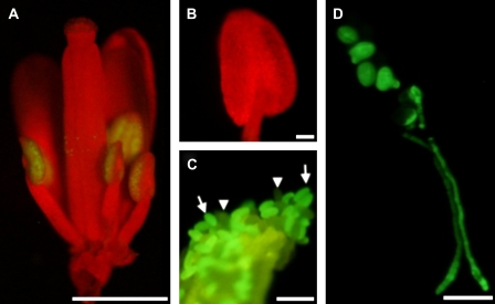
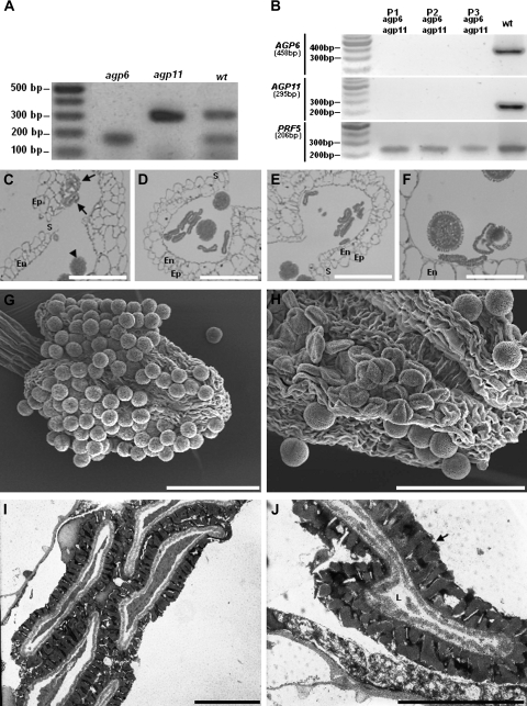
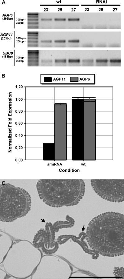
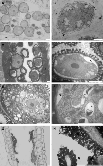
Arabinogalactan proteins (AGPs) are structurally complex plasma membrane and cell wall proteoglycans that are implicated in diverse developmental processes, including plant sexual reproduction. Male gametogenesis (pollen grain development) is fundamental to plant sexual reproduction. The role of two abundant, pollen-specific AGPs, AGP6, and AGP11, have been investigated here. The pollen specificity of these proteoglycans suggested that they are integral to pollen biogenesis and their strong sequence homology indicated a potential for overlapping function. Indeed, single gene transposon insertion knockouts for both AGPs showed no discernible phenotype. However, in plants homozygous for one of the insertions and heterozygous for the other, in homozygous double mutants, and in RNAi and amiRNA transgenic plants that were down-regulated for both genes, many pollen grains failed to develop normally, leading to their collapse. The microscopic observations of these aborted pollen grains showed a condensed cytoplasm, membrane blebbing and the presence of small lytic vacuoles. Later in development, the generative cells that arise from mitotic divisions were not seen to go into the second mitosis. Anther wall development, the establishment of the endothecium thickenings, the opening of the stomium, and the deposition of the pollen coat were all normal in the knockout and knockdown lines. Our data provide strong evidence that these two proteoglycans have overlapping and important functions in gametophytic pollen grain development.


Figures

Fig. 1.
Fig. 1.
Transgenic Arabidopsis plants expressing a ProAGP6:GFP construct. (A) Low-power binocular fluorescence microscope of a flower in a stage where the anthers have their locules already formed and are extending. GFP fluorescence is exclusively visible in pollen grains. (B) Anther at the immediately preceding stage of development, with reference to the stage in (A). No fluorescence could be observed. Typically in this stage the locules are formed and microspores are released from tetrads. (C) Mature dehiscent anther, showing GFP-labelled (arrows) and unlabelled pollen grains (arrow heads). The anther wall is also visible as green-yellow autofluorescence. (D) Fluorescence microscopy of germinating mature pollen. GFP fluorescence is visible in all pollen grains and along all pollen tube extension. Bars: (A) 1 mm; (B, C) 500 μm; (D) 50 μm. (This figure is available in colour at JXB online.)
Fig. 2.
Fig. 2.
Ds transposon tag lines for AGP6 and AGP11. (A) Duplex RT-PCR amplification products of AGP6 and AGP11 mRNA transcripts in pollen of Arabidopsis wild-type (wt), and of Arabidopsis tag lines containing the Ds transposon in AGP6 (agp6) and in AGP11 (agp11) genes. Only in wild-type plants are the two expected amplification products visible. (B) RT-PCR amplification products of AGP6 and AGP11 mRNA transcripts in pollen of wild-type Arabidopsis (wt), and of three agp6 agp11 double mutant plants (P1, P2, P3). Figures under AGP and reference gene names refer to expected sizes of PCR amplification products. (C) Light micrograph of an anther of F1 plants, heterozygous for AGP6 and AGP11. Pollen grains show a collapsing phenotype (arrows) that contrasts with the ones that are roughly spherical pollen grains, and show no phenotype (arrow head). (D) Light micrograph of an anther of an agp6 F2 plant (and heterozygous for AGP11). At the time of dehiscence, more than 50% of the pollen grains show a collapsed phenotype. (E) Light micrograph of an anther of an agp11 F2 plant (and heterozygous for AGP6), exhibiting the same pollen morphology shown in (D). (F) Light micrograph of an anther of an agp6 agp11 double mutant F3 plant. Collapsing of pollen grains is evident, while some pollen grains still show a normal morphology. (G) Scanning electron micrograph of a wild-type dehiscent anther, showing normal roundish pollen grains. (H) Scanning electron micrograph of an agp6 agp11 double mutant dehiscent anther. The collapsed pollen grains are evident. (I) Transmission electron micrograph of an agp6 agp11 double mutant. The collapse of the pollen grain is evident. (J) High magnification transmission electron micrograph of pollen grains from of an agp6 agp11 double mutant. The pollen grain in the image shows a reduced cell lumen and a well-developed exine wall (arrow). En, endothecium; Ep, epidermis; L, cell lumen; S, stomium. Bars: (C, D, E, F) 20 μm; (G, H) 60 μm; (I, J) 5 μm.
Fig. 3.
Fig. 3.
Down-regulation of AGP6 and AGP11 in RNAi and amiRNA plants. (A) RT-PCR amplification products of AGP6 and AGP11 mRNA transcripts in mature anthers of wild-type (wt) and RNAi Arabidopsis. Figures in the top row refer to PCR cycle numbers. UBC9 was used as the reference gene. (B) Real-time RT-PCR amplification products of AGP6 and AGP11 mRNA transcripts in anthers of wild-type (wt) and amiRNA Arabidopsis. In the panel each bar represents an average of two independent reactions and technical replicates. The anthers were at stage 10 of pollen development according to Smyth et al. (1990). AGP6 and AGP11 transcript levels were normalized to UBC9 levels. (C) Light micrograph of an anther from a plant exhibiting an amiRNA construction. Some pollen grains show a collapsing phenotype (arrow) while others look phenotypically normal. Bar: 20 μm.
Fig. 4.
Fig. 4.
Microscopy images of plants down-regulated for AGP6 and AGP11 by RNA interference (RNAi). (A) Light micrograph of an anther from a wild-type plant showing normal pollen morphology at the stage of mature microspores. (B) Electron micrograph of an anther from an RNAi plant at the tetrad stage of development. The young microspore, surrounded by the callose wall, shows the initiation of the building of the exine wall (arrow). (C) Light micrograph of an anther from an RNAi plant at the beginning of the microspore stage of development. The anther shows a well-developed tapetum. As expected for this stage of development the endothecium is not differentiated yet. (D) Electron micrograph of a pollen grain from the anther in (C); the retraction of the plasma membrane is evident (arrow). The exine wall shows its final architecture. (E) Electron micrograph of a wild-type pollen grain. (F) Higher magnification of one of the pollen grains in (C), showing the presence of lytic vacuoles (*) as well as the cytoplasm retraction (arrow). (G) Light micrograph of an anther from an RNAi plant at the end of pollen development. It is evident the collapsing of all the pollen grains observed inside the pollen sac. (H) Electron micrograph detail from the same anther in (G). The anther releases empty pollen grains completely devoid of contents, with a well-developed pollen coat (arrow). Ep, epidermis; En, endothecium; T, tapetum. Bars: 5 μm.

References

    1. Acosta-Garcia G, Vielle-Calzada JP. A classical arabinogalactan protein is essential for the initiation of female gametogenesis in Arabidopsis. The Plant Cell. 2004;16:2614–2628. - PMC - PubMed
    1. Alfieiri JA, Martin AD, Takeda J, Kondoh G, Myles DG, Primakoff P. Infertility in female mice with an oocyte-specific knockout of GPI-anchored proteins. Journal of Cell Science. 2003;116:2149–2155. - PubMed
    1. Cheung AY, Wang H, Wu HM. A floral transmitting tissue-specific glycoprotein attracts pollen tubes and stimulates their growth. Cell. 1995;82:383–393. - PubMed
    1. Clough SJ, Bent AF. Floral dip: a simplified method for Agrobacterium-mediated transformation of Arabidopsis thaliana. The Plant Journal. 1998;16:735–743. - PubMed
    1. Coimbra S, Almeida J, Junqueira V, Costa M, Pereira LG. Arabinogalactan proteins as molecular markers in Arabidopsis thaliana sexual reproduction. Journal of Experimental Botany. 2007;58:4027–4035. - PubMed

Publication types