Review
. 2009 Dec;82(984):1034-42.
doi: 10.1259/bjr/99354802. Epub 2009 May 11.

Mullerian duct anomalies: from diagnosis to intervention

Affiliations
Review

Mullerian duct anomalies: from diagnosis to intervention

T M Chandler et al. Br J Radiol. 2009 Dec.

Abstract

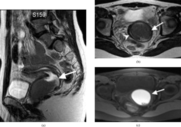
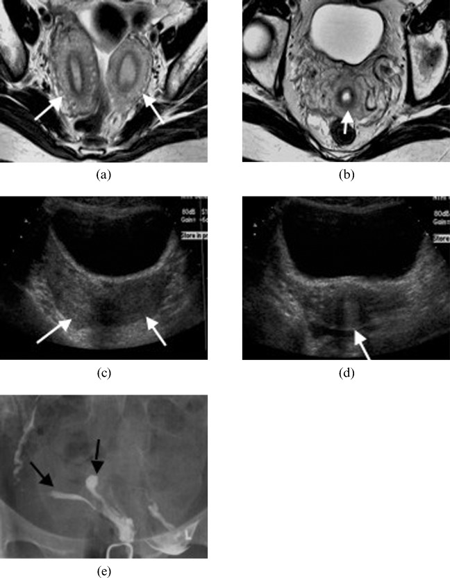
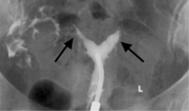
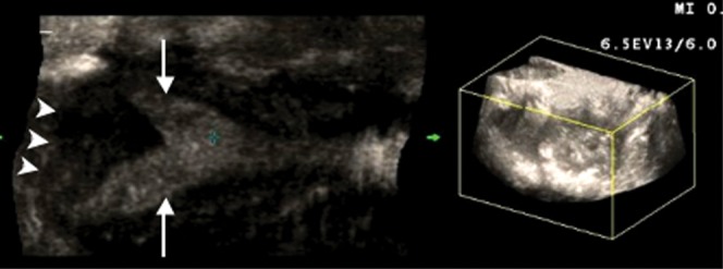
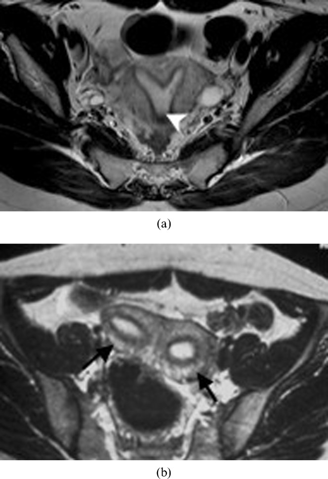
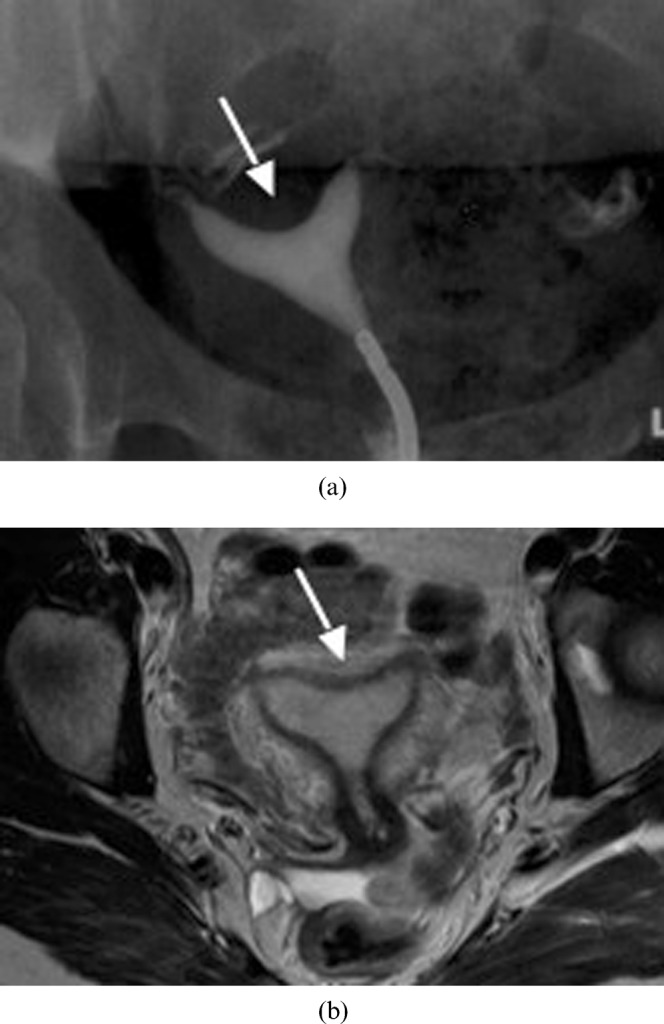
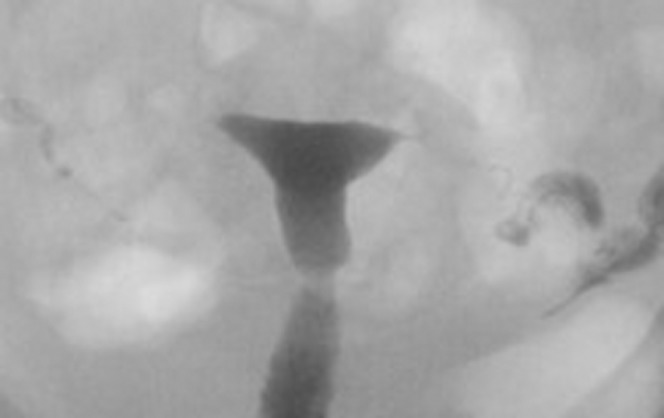
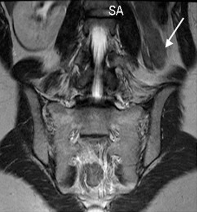
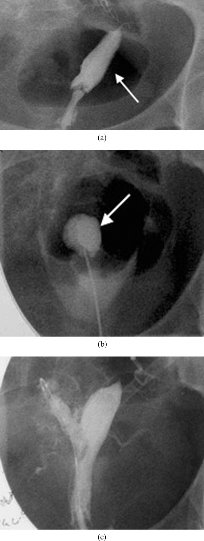
The purpose of this study was to review the embryology, classification, imaging features and treatment options of Müllerian duct anomalies. The three embryological phases will be described and the appearance of the seven classes of Müllerian duct anomalies will be illustrated using hysterosalpingography, ultrasound and MRI. This exhibit will also review the treatment options, including interventional therapy. The role of imaging is to help detect, classify and guide surgical management. At this time, MRI is the modality of choice because of its high accuracy in detecting and accurately characterising Müllerian duct anomalies. In conclusion, radiologists should be familiar with the imaging features of the seven classes of Müllerian duct anomalies, as the appropriate course of treatment relies upon the correct diagnosis and categorisation of each anomaly.


Figures

Figure 1.
Figure 1.
The classification system of Müllerian duct anomalies used by the American Fertility Society.
Figure 2.
Figure 2.
A 19 year-old woman with uterine and vaginal agenesis (Class I). (a,b) Axial and (c) sagittal T2 MRI of the pelvis demonstrates normal ovaries (arrow depicts the right ovary; left not shown) and absent uterus and upper vagina consistent with Mayer–Rokitansky–Kuster–Hauser syndrome. Note the ovaries are not visualised in (b,c).
Figure 3.
Figure 3.
A 22-year-old woman with a unicornuate uterus with no rudimentary horn (Class II). Axial T2 weighted MRI illustrates the classic banana shape appearance of the unicornuate uterus (arrow).
Figure 4.
Figure 4.
A 29-year-old woman with a unicornuate uterus and a rudimentary horn (Class II). (a) Hysterosalpingogram reveals a unicornuate uterus (arrow) with a rudimentary communicating horn (arrowhead). (b) Correlative axial T2 weighted MRI confirms that there is no obstructing component beyond the patent rudimentary horn (arrowhead).
Figure 5.
Figure 5.
A 26-year-old woman with uterus didelphys (Class III). (a) Axial T2 weighted MRI demonstrating complete duplication of the uterine horns (arrows) and cervices (arrowheads). (b) A septum (arrow) dividing two vaginal canals.
Figure 6.
Figure 6.
A 19-year-old woman with didelphys uterus (Class III). (a) Sagittal T2 weighted MR image of a didelphys uterus with a transverse vaginal septum causing unilateral haematocolpos (arrow). (b) Axial T2 weighted MR image shows a dilated left vaginal canal with predominantly low signal intensity fluid (arrow) and compressed right vaginal canal (arrowhead). (c) Correlating axial T1 weighted fat saturation MR image shows hyperintense fluid in the distended left vaginal canal in keeping with haematocolpos (arrow).
Figure 7.
Figure 7.
A 30-year-old woman with a bicornuate uterus (Class IV). Axial T2 weighted image illustrating (a) two separate uterine horns (arrows) that fuse at its inferior end to give (b) a single cervix (arrow). Transverse ultrasound images demonstrating (c) two uterine horns (arrows) and (d) a single cervix (arrow). (e) Hysterosalpingography of a bicornuate uterus. The contrast material fills two separate uterine horns (arrows).
Figure 8.
Figure 8.
A 32-year-old woman with a septate uterus (Class V). Hysterosalpingography demonstrates that there are two uterine horns (arrows).
Figure 9.
Figure 9.
A 30-year-old woman with a septate uterus (Class V). A two-dimensional coronal reconstruction (left) of the three-dimensional volume image (right) shows two uterine horns (arrows) and a flat fundal contour (arrowheads).
Figure 10.
Figure 10.
A 25-year-old woman with a septate uterus (Class V) who underwent a spontaneous abortion. (a,b) Subsequent MRI performed on this patient confirms the septate uterus. Axial T2 weighted image shows two uterine horns (arrows) with incomplete resorption of the midline septum (arrowhead) (a). A faint fibrous segment is seen between the two horns (b).
Figure 11.
Figure 11.
Arcuate uterus (Class VI). (a) Hysterosalpingogram of a 36-year-old woman with an arcuate uterus. Note the mild indentation of the endometrium at the uterine fundus (arrow). (b) Axial T2 weighted MR image of an arcuate uterus in a 28-year-old woman. Note the mild curvature at the fundus (arrow).
Figure 12.
Figure 12.
A 30-year-old woman with diethylstilbestrol exposure in utero (Class VII). Hysterosalpingography illustrates the T-shaped uterus.
Figure 13.
Figure 13.
A 28-year-old woman with unicornuate uterus and a renal anomaly. This coronal MR image of the retroperitoneum illustrates a congenitally absent left kidney (arrow) in a patient known to have a unicornuate uterus.
Figure 14.
Figure 14.
A 22-year-old with intense menstrual pain who was investigated and found to have a bicornuate uterus with a non-communicating right horn. (a) Hysterosalpingogram reveals filling of the left horn (arrow) but not the obstructed right horn. (b) Intraoperative hystosalpingogram illustrating the puncture through the uterine septum into the obstructed right horn (arrow). This was performed following needle puncture of the septum with ultrasound guidance. A wire was then placed into the cavity and the gynaecologist then used the wire to guide resection of part of the wall of the obstructing right horn. (c) Subsequent hysterosalpingogram shows filling of the previously obstructed right horn. Note that spasm of the right tube prevented uniform tubal filling. Subsequent selective salpingography demonstrated the right tube to be patent. The patient had resolution of her pain and was very pleased at 2 year follow-up.

References

    1. The American Fertility Society classifications of adnexal adhesions, distal tubal obstruction, tubal occlusions secondary to tubal ligation, tubal pregnancies, Müllerian anomalies and intrauterine adhesions. Fertil Steril 1998;49:944–55 - PubMed
    1. Deutch TD, Abuhamad AZ. The role of 3-dimensional ultrasonography and magnetic resonance imaging in the diagnosis of Müllerian duct anomalies. J Ultrasound Med 2008;27:413–23 - PubMed
    1. Mueller GC, Hussain HK, Smith YR, Quint EH, Carlos RC, Johnson TD, et al. Müllerian duct anomalies: comparison of MRI diagnosis and clinical diagnosis. AJR Am J Roentgenol. 2007;189:1294–302 - PubMed
    1. Heinonen PK, Saarikoski S, Pystynen P. Reproductive performance of women with uterine anomalies: an evaluation of 182 cases. Acta Obstet Gynecol Scand 1982;61:157–62 - PubMed
    1. Homer HA, Li TC, Cooke ID. The septate uterus: a review of management and reproductive outcome. Fertil Steril 2000;73:1–14 - PubMed