. 2009 Jul;37(12):4127-34.
doi: 10.1093/nar/gkp340. Epub 2009 May 11.

The formation pathway of i-motif tetramers

Affiliations

The formation pathway of i-motif tetramers

Jean-Louis Leroy. Nucleic Acids Res. 2009 Jul.

Abstract

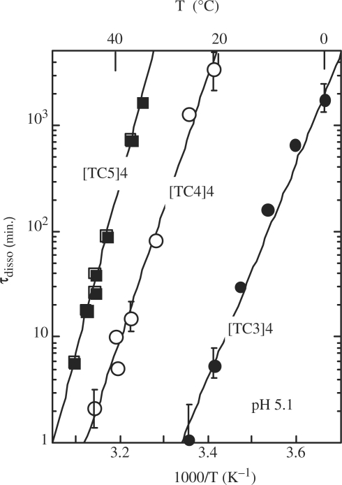
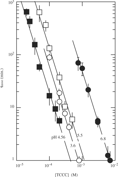
The i-motif is a four-stranded structure formed by two intercalated parallel duplexes containing hemiprotonated C*C(+) pairs. In order to describe the sequence of reactions by which four C-rich strands associate, we measured the formation and dissociation rates of three [TC(n)](4) tetramers (n = 3, 4 and 5), their dissociation constant and the reaction order for tetramer formation by NMR. We find that TC(n) association results in the formation of several tetramers differing by the number of intercalated C*C(+) pairs. The formation rates of the fully and partially intercalated species are comparable but their lifetimes increase strongly with the number of intercalated C*C(+) pairs, and for this reason the single tetramer detected at equilibrium is that with optimal intercalation. The tetramer half formation times vary as the power -2 of the oligonucleotide concentration indicating that the reaction order for i-motif formation is 3. This observation is inconsistent with a model supposing association of two preformed duplex and suggests that quadruplex formation proceeds via sequential strand association into duplex and triplex intermediate species and that triplex formation is rate limiting.


Figures

Figure 1.
Figure 1.
Lifetime versus temperature at pH 5.1 of the fully intercalated i-motif tetramers of [TC3]4 (black circles), [TC4]4 (open circles) and [TC5]4 (black squares). The lifetime of [TC2]4 is estimated in the range of 3 s to 3 min at 0°C.
Figure 2.
Figure 2.
Effect of pH on the formation and dissociation times and on the stability of the fully intercalated [TC3]4 tetramer. (A) [TC3]4 lifetime (open circles) and half formation time in 10−4 M TC3 solutions (black circles). The half formation time varies with pH as the power −2 of the product of the fraction of neutral cytidine, formula image and protonated cytidine: formula image
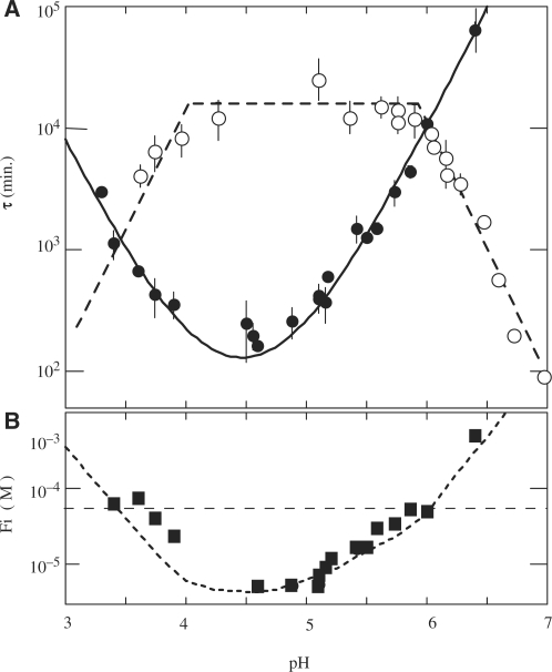
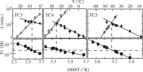
. (B) Reduced dissociation constant versus pH. The observation that Fi is close 10−4/2 (dashed line) at the temperature for which the association time measured in 10−4 M TC3 solution and the dissociation time are equal shows the consistence of the kinetics and equilibrium measurements.
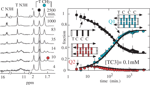
Figure 3.
Figure 3.
Association kinetics of TC3 at 0°C pH 5.1 into i-motif structures. The proton spectra of a 10−4 M TC3 solution initially monomeric recorded as a function of the time (left panel) shows the formation of two multimers. The imino protons and the methyl peaks of the fully intercalated tetramer are labeled by blue dots and the methyl peak of the monomer by a black dot. The spectra show the transient accumulations of a short-lived species at the beginning of the kinetics (red dot), which is tentatively identified as the incompletely intercalated structure displayed on the figure. The solid line fitting the time evolution of the monomer and tetramer fractions are computed according to a model involving two parallel reactions (cf. text). The simulation indicates that the formation rate of both tetramers is comparable: 1/60 min and 1/90 min for the red and blue species, respectively, but their lifetimes are extremely different: 5 min and 1900 min for the red and blue species, respectively.
Figure 4.
Figure 4.
Half formation time of the fully intercalated [TC3]4 tetramer versus the oligonucleotide concentration at 0°C pH 6.8 (black circles), 5.5 (open squares), 4.56 (black squares) and 3.6 (open circles). The lines drawn through the data points show that the half reaction time increases as power of −2 of the oligonucleotide concentration and therefore establish that the reaction order is 3.
Figure 5.
Figure 5.
Effect of temperature on the formation and dissociation times and on the stability of [TC3]4 (left), [TC4]4 (center) and [TC5]4 (right) at pH 5.1. Upper panels: half formation times (open circles) and dissociation times (black circle) of the fully intercalated tetramers. The half formation times were measured in 10−4 M oligonucleotide solutions or extrapolated at 10−4 M from values measured at higher oligonucleotide concentrations. Lower panels: reduced dissociation constants derived from the monomer and tetramer fractions at equilibrium (black squares).
Figure 6.
Figure 6.
Association kinetics of TC4 into i-motif structures in 1.2 mM solution at 0°C, pH 5.1. (A) The imino proton region of spectra collected as a function of the time shows the formation of three tetramers. The markers of the fully intercalated tetramer are labeled by green circles. The imino protons peaks labeled by blue circles are tentatively assigned to the species with an empty intercalation site between T1 and C2 and those labeled by red dot to the species whose T1-C2 and C2-C3 intercalation sites are empty. (B) Evolution of the fractions of each species as a function of the time. The monomer fraction (black circle) was determined by the intensity of monomer methyl peak. The full lines fitting the evolution of each species were computed according to a system of parallel reaction between the monomer and each tetramer. The tetramer formation and dissociation times used in the simulation are displayed in Figure 7.
Figure 7.
Figure 7.
Formation (A) and dissociation times (B) of the fully and partially intercalated tetramer of TC4 derived from the fit of the evolution of the different species in 1.2 mM TC4 solution at 0°C. The color code is the same than in Figure 6. The tetramer formation times are nearly independent of the intercalation topology. The heavy dashed line shows the lifetimes measured (Figure 1) for the fully intercalated TC4 tetramer.

References

    1. Gehring K, Leroy J-L, Guéron M. A tetrameric DNA structure with protonated cytidine-cytidine base pairs. Nature. 1993;363:561–565. - PubMed
    1. Patel D, Bouaziz S, Kettani A, Wang Y. Structure of guanine-rich and cytosine-rich quadruplexes formed in vitro by telomeric, centomeric and triplet repeat disease DNA sequences. In: Neidle S, editor. Oxford Handbook of Nucleic Acid Structure. Oxford University Press; 1999. pp. 389–454.
    1. Cai L, Chen L, Raghavan S, Ratliff R, Moyzis R, Rich A. Intercalated cytosine motif and novel adenine clusters in the crystal structure of the tetrahymena telomere. Nucleic Acids Res. 1998;26:4696–4705. - PMC - PubMed
    1. Leroy JL, Guéron M. Solution structure of the i-motif tetramers of d(TCC), d(5methylCCT) and d(T5methylCC): novel NOE connection between amino protons and sugar protons. Structure. 1995;3:101–120. - PubMed
    1. Nonin S, Phan AT, Leroy J-L. Solution structure and base-pair opening kinetics of the i-motif dimer of d(5mCCTTTACC): a non canonical structure with possible role in chromosome stability. Structure. 1997;5:1231–1246. - PubMed

Publication types