. 2009 Sep;23(9):3226-32.
doi: 10.1096/fj.09-132019. Epub 2009 May 11.

Deletion of the secretory vesicle proteins IA-2 and IA-2beta disrupts circadian rhythms of cardiovascular and physical activity

Affiliations

Deletion of the secretory vesicle proteins IA-2 and IA-2beta disrupts circadian rhythms of cardiovascular and physical activity

Soo Mi Kim et al. FASEB J. 2009 Sep.

Abstract

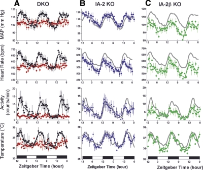
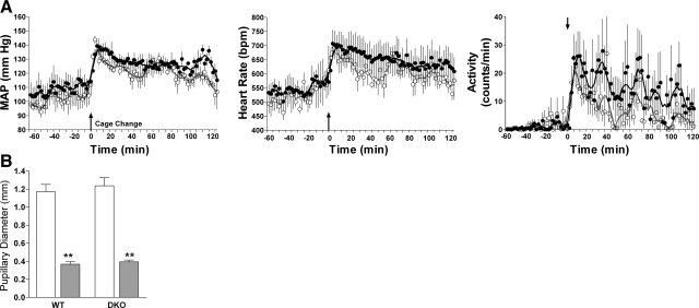
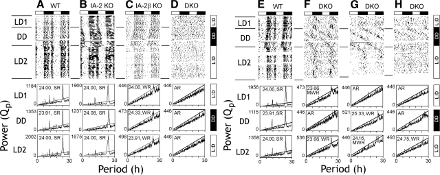
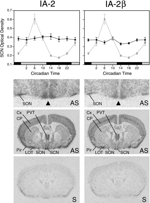
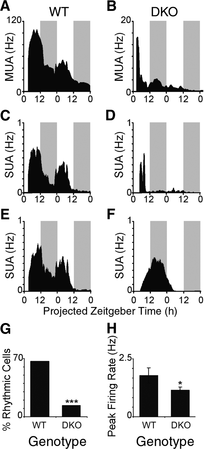
Targeted deletion of IA-2 and IA-2beta, major autoantigens in type 1 diabetes and transmembrane secretory vesicle proteins, results in impaired secretion of hormones and neurotransmitters. In the present study, we evaluated the effect of these deletions on daily rhythms in blood pressure, heart rate, core body temperature, and spontaneous physical and neuronal activity. We found that deletion of both IA-2 and IA-2beta profoundly disrupts the usual diurnal variation of each of these parameters, whereas the deletion of either IA-2 or IA-2beta alone did not produce a major change. In situ hybridization revealed that IA-2 and IA-2beta transcripts are highly but nonrhythmically expressed in the suprachiasmatic nuclei, the site of the brain's master circadian oscillator. Electrophysiological studies on tissue slices from the suprachiasmatic nuclei showed that disruption of both IA-2 and IA-2beta results in significant alterations in neuronal firing. From these studies, we concluded that deletion of IA-2 and IA-2beta, structural proteins of secretory vesicles and modulators of neuroendocrine secretion, has a profound effect on the circadian system.


Figures

Figure 1.
Figure 1.
Mean arterial pressure (MAP), heart rate, spontaneous physical activity, and body temperature in WT and KO mice. A) WT (black) and DKO mice (red). B) IA-2 KO mice (blue). C) IA-2β KO mice (green). Gray lines in B and C represent WT data from A. Data represent means ± se of 5 to 8 mice/group, where the value for each mouse is the hourly average of 10-s recordings taken every 2 min. Mice were maintained in a 12-h light-dark (LD) lighting cycle, and data were collected continuously for 60 h. Zeitgeber times 0 and 12 correspond to lights on at 6:00 AM and lights off at 6:00 PM, respectively. Lines represent data smoothing using the weighted average of the 9 nearest points.
Figure 2.
Figure 2.
A) Effect of acute stress induced by cage change on MAP, heart rate, and spontaneous physical activity in WT (•) and DKO (○) mice. At time 0, mice were transferred into cages previously occupied by different animals. Points represent 10-s data collections taken every 2 min. Vertical lines represent se of 3 to 5 mice/group. B) Effect of light exposure [transition from dark (□) to medium bright light (▒); 1.2 mW cm−2] on pupillary diameter of WT and DKO mice. Vertical lines represent se of 3 to 5 mice/group.
Figure 3.
Figure 3.
Wheel-running rhythms of WT, IA-2-KO, IA-2β-KO, and DKO mice. Double-plotted actograms (top panels) and χ2 periodograms (bottom panels) show activity under light-dark (LD1), constant dark (DD), and LD2 conditions. A, E) WT mice. B) IA-2 KO mouse. C) IA-2β KO mouse. D, F–H) IA-2/IA-2β DKO mice. Actograms depict wheel rotations double-plotted over 48 h. Rhythm strength (Qp) is shown on the ordinate and period on the abscissa of the periodograms. Peak Qp value for each panel is shown. Within each panel, period estimate and strength of rhythmicity are indicated. Components traversing the solid diagonal line in each panel are significant at P < 0.05 (χ2 periodogram). DKO mice express a variety of behavioral phenotypes, with some evidence of a weak rhythmic component when released back into LD (LD2). SR, strong rhythm; WR, single weak rhythm; MWR, multiple weak rhythms; AR, arrhythmic.
Figure 4.
Figure 4.
Expression of IA-2 (left) and IA-2β (right) (dark black lines) as determined by in situ hybridization in brain sections through the SCN from WT mice housed in constant darkness (top panels). IA-2 and IA-2β showed no evidence of rhythmicity (P>0.05; ANOVA,). Adjacent sections from the same animals revealed the expected high-amplitude rhythm of mPer1 expression (light gray symbols with same line plotted in both left and right panels). Values represent means ± se of 5 to 6 animals. Sections hybridized with antisense (AS) probes showed that both IA-2 and IA-2β genes are highly expressed in the SCN, with no obvious subregional localization. Top image is a higher magnification of the image below it. Triangle indicates the bilaterally symmetrical SCN. Sense (S) strand control probes showed the level of the background hybridization (bottom panels). Cx, cortex; CP, caudate-putamen; LOT, nucleus of the lateral olfactory tract; Pir, piriform cortex; SON, supraoptic nucleus; PVT, paraventricular nucleus of the thalamus.
Figure 5.
Figure 5.
Neuronal firing rate rhythms are disrupted in the SCN of DKO mice. WT mice show clear rhythms in MUA (A) and SUA (B, C), whereas DKO mice show blunted or arrhythmic MUA (D) and SUA profiles (E, F). Firing rate traces show the average firing rate each minute from a representative slice, smoothed using a 1-h moving average. Shaded gray bars represent the projected lights-off period. In contrast to the neurons (n=15) from the WT mice, the percentage of rhythmic neurons (G) and the peak firing rate (H) of neurons (n=22) from DKO mice are significantly reduced. *P < 0.05; unpaired t test. ***P < 0.001; χ2 test.

References

    1. Dunlap J C, Loros J J, DeCoursey P J. Sunderland, MA, USA: Sinauer Associates; ChronobiologyBiological Timekeeping. 2004
    1. Reppert S M, Moore R Y, Klein D C. New York.: Oxford University Press; Suprachiasmatic NucleusThe Mind’s Clock. 1991
    1. Brown T M, Piggins H D. Electrophysiology of the suprachiasmatic circadian clock. Prog Neurobiol. 2007;82:229–255. - PubMed
    1. Reppert S M, Weaver D R. Coordination of circadian timing in mammals. Nature. 2002;418:935–941. - PubMed
    1. Aton S J, Colwell C S, Harmar A J, Waschek J, Herzog E D. Vasoactive intestinal polypeptide mediates circadian rhythmicity and synchrony in mammalian clock neurons. Nat Neurosci. 2005;8:476–483. - PMC - PubMed

Publication types

MeSH terms

Substances

LinkOut - more resources