. 2009 May;66(5):620-31.
doi: 10.1001/archneurol.2009.48.

The common inhalational anesthetic sevoflurane induces apoptosis and increases beta-amyloid protein levels

Affiliations

The common inhalational anesthetic sevoflurane induces apoptosis and increases beta-amyloid protein levels

Yuanlin Dong et al. Arch Neurol. 2009 May.

Abstract

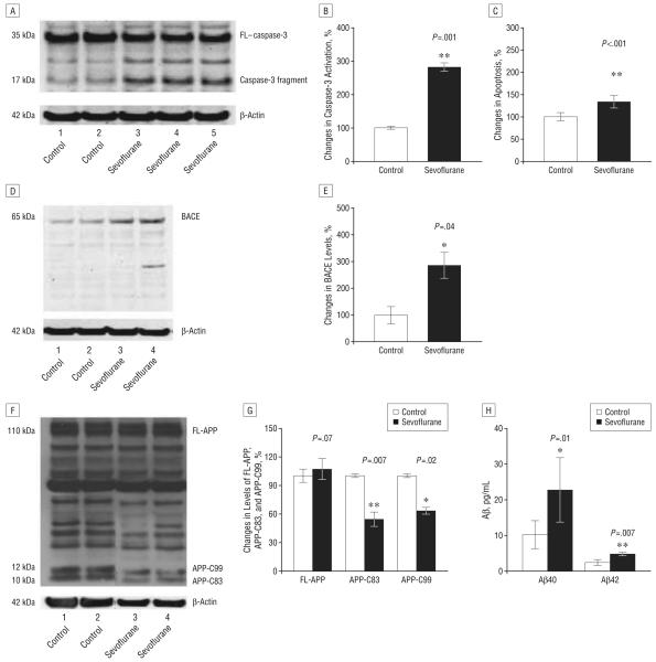
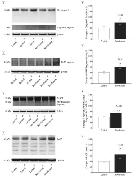
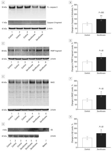
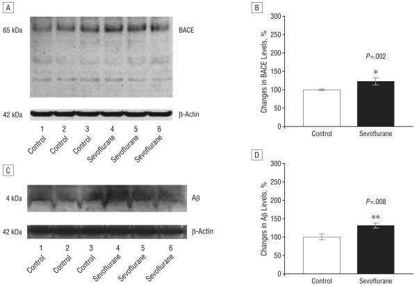
Objective: To assess the effects of sevoflurane, the most commonly used inhalation anesthetic, on apoptosis and beta-amyloid protein (Abeta) levels in vitro and in vivo. Subjects Naive mice, H4 human neuroglioma cells, and H4 human neuroglioma cells stably transfected to express full-length amyloid precursor protein.

Interventions: Human H4 neuroglioma cells stably transfected to express full-length amyloid precursor protein were exposed to 4.1% sevoflurane for 6 hours. Mice received 2.5% sevoflurane for 2 hours. Caspase-3 activation, apoptosis, and Abeta levels were assessed.

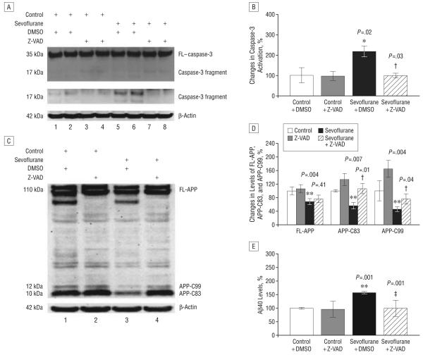
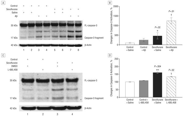
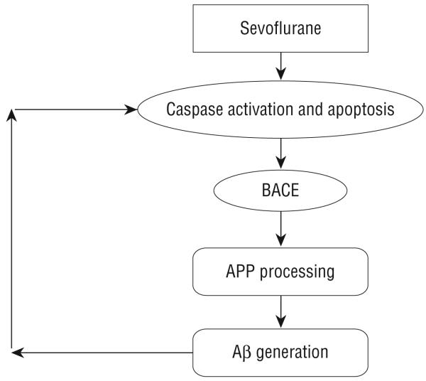
Results: Sevoflurane induced apoptosis and elevated levels of beta-site amyloid precursor protein-cleaving enzyme and Abeta in vitro and in vivo. The caspase inhibitor Z-VAD decreased the effects of sevoflurane on apoptosis and Abeta. Sevoflurane-induced caspase-3 activation was attenuated by the gamma-secretase inhibitor L-685,458 and was potentiated by Abeta. These results suggest that sevoflurane induces caspase activation which, in turn, enhances beta-site amyloid precursor protein-cleaving enzyme and Abeta levels. Increased Abeta levels then induce further rounds of apoptosis.

Conclusions: These results suggest that inhalational anesthetic sevoflurane may promote Alzheimer disease neuropathogenesis. If confirmed in human subjects, it may be prudent to caution against the use of sevoflurane as an anesthetic, especially in those suspected of possessing excessive levels of cerebral Abeta.


Figures

Figure 1
Figure 1
Treatment with 4.1% sevoflurane for 6 hours induces caspase-3 activation and apoptosis, alters amyloid precursor protein (APP) processing, and increases levels of β-site APP-cleaving enzyme (BACE) and β-amyloid protein (Aβ) in H4-APP cells. A, Sevoflurane treatment induces caspase-3 cleavage (activation) compared with control conditions in H4-APP cells in Western blot analysis; there is no significant difference in the amounts of β-actin between the control conditions and the sevoflurane-treated H4-APP cells. B, Caspase-3 activation was assessed by quantifying the ratio of caspase-3 fragment to full-length (FL)–caspase-3 in Western blot analysis. Quantification of the Western blot shows that sevoflurane treatment increases caspase-3 activation compared with control conditions, normalized to β-actin levels. C, Sevoflurane treatment increases apoptosis compared with control conditions in H4-APP cells. D, Sevoflurane treatment enhances levels of BACE compared with control conditions in H4-APP cells in Western blot analysis; there is no significant difference in the amounts of β-actin in the control conditions or the sevoflurane-treated H4-APP cells. E, Quantification of the Western blot shows that sevoflurane treatment enhances BACE levels compared with control conditions in H4-APP cells, normalized to β-actin levels. F, Sevoflurane treatment decreases levels of APP-C83 and APP-C99 compared with control conditions in Western blot analysis; there is no significant difference in the amounts of β-actin in the control conditions or the sevoflurane-treated H4-APP cells. G, Quantification of the Western blot shows that sevoflurane treatment decreases levels of APP-C83 and APP-C99, but not FL-APP, compared with control conditions in H4-APP cells, normalized to β-actin levels. H, Sevoflurane treatment increases levels of both Aβ40 and Aβ42 compared with control conditions. Data are mean (SD); n=3 to 10 for each experimental group; the t test was used to compare the difference between control conditions and sevoflurane treatment conditions; values are significant at *P<.05 and **P<.01.
Figure 2
Figure 2
Anesthesia with 2.5% sevoflurane for 2 hours induces caspase-3 activation and increases β-site amyloid precursor protein (APP)–cleaving enzyme (BACE) levels 6 hours after anesthesia in naive mice. A, Sevoflurane anesthesia induces caspase-3 cleavage (activation) by decreasing full-length (FL)–caspase-3 levels and increasing caspase-3 fragment compared with control conditions in Western blot analysis. B, Quantification of the Western blots shows that sevoflurane anesthesia increases the ratio of caspase-3 fragment to FL–caspase-3 levels compared with control conditions. C, Sevoflurane anesthesia induces poly–(adenosine diphosphate–ribose) polymerase (PARP) cleavage by increasing PARP fragment compared with control conditions in Western blot analysis. D, Quantification of the Western blots shows that sevoflurane anesthesia increases levels of PARP fragment compared with control conditions. E, Sevoflurane anesthesia increases levels of caspase-cleaved APP-N-fragment (APP-N-caspase fragment) compared with control conditions in Western blot analysis. F, Quantification of the Western blots shows that sevoflurane anesthesia increases levels of APP-N-caspase fragment compared with control conditions. G, Sevoflurane anesthesia increases BACE levels compared with control conditions in Western blot analysis. H, Quantification of the Western blots shows that sevoflurane anesthesia increases BACE levels compared with control conditions. There is no significant difference in amounts of β-actin in control conditions or sevoflurane-treated mouse brain tissue. Data are means (SD); n=3 to 6 for each experimental group; the t test was used to compare the difference between control conditions and sevoflurane anesthesia; values are significant at * P<.05 and ** P<.01.
Figure 3
Figure 3
Anesthesia with 2.5% sevoflurane for 2 hours induces caspase activation and increases levels of β-site amyloid precursor protein (APP)–cleaving enzyme (BACE) and β-amyloid protein (Aβ) 12 hours after anesthesia. A, Sevoflurane anesthesia induces caspase-3 cleavage (activation) by decreasing full-length (FL)–caspase-3 levels and increasing caspase-3 fragment compared with control conditions in Western blot analysis. B, Quantification of the Western blots shows that sevoflurane anesthesia increases the ratio of caspase-3 fragment to FL–caspase-3 levels compared with control conditions. C, Sevoflurane anesthesia induces poly–(adenosine diphosphate–ribose) polymerase (PARP) cleavage by increasing PARP fragment compared with control conditions in Western blot analysis. D, Quantification of the Western blots shows that sevoflurane anesthesia increases levels of PARP fragment compared with control conditions. E, Sevoflurane anesthesia increases BACE levels compared with control conditions in Western blot analysis. F, Quantification of the Western blot shows that sevoflurane anesthesia increases BACE levels compared with control conditions. G, Sevoflurane anesthesia increases Aβ levels compared with control conditions in Western blot analysis. Synthetic Aβ was used as a marker to identify position of Aβ in Western blot analysis. H, Quantification of the Western blot shows that sevoflurane anesthesia increases Aβ levels compared with control conditions. There is no significant difference in amounts of β-actin in control conditions or sevoflurane-treated mouse brain tissue. Data are mean (SD); n=3 to 6 for each experimental group; the t test was used to compare the difference between control conditions and sevoflurane anesthesia; values are significant at *P<.05 and **P<.01.
Figure 4
Figure 4
Anesthesia with 2.5% sevoflurane for 2 hours increases levels of β-site amyloid precursor protein–cleaving enzyme (BACE) and β-amyloid protein (Aβ) 24 hours following anesthesia. A, Sevoflurane anesthesia increases BACE levels compared with control conditions in Western blot analysis. B, Quantification of the Western blot shows that sevoflurane anesthesia increases BACE levels compared with control conditions. C, Sevoflurane anesthesia increases Aβ levels compared with control conditions in Western blot analysis. D, Quantification of the Western blot shows that sevoflurane anesthesia increases Aβ levels compared with control conditions. There is no significant difference in amounts of β-actin in control conditions or sevoflurane-treated mouse brain tissue. Data are mean (SD); n=3 to 6 for each experimental group; the t test was used to compare the difference between control conditions and sevoflurane anesthesia; values are significant at *P<.05 and **P<.01.
Figure 5
Figure 5
The caspase inhibitor Z-VAD inhibits caspase-3 activation and attenuates sevoflurane-induced increases in β-amyloid protein (Aβ) in H4 amyloid precursor protein (H4-APP) cells. A, Western blot analysis shows that 4.1% sevoflurane induces caspase-3 cleavage (activation) compared with control conditions or Z-VAD (100μM) treatment. The Z-VAD treatment inhibits the caspase-3 cleavage (activation) induced by 4.1% sevoflurane treatment. The blot showing the band of caspase-3 fragment only is the same blot with more exposure time in developing the film. There is no significant difference in the amounts of β-actin in H4-APP cells with the above treatments. B, Quantification of the Western blot shows that sevoflurane treatment increases caspase-3 activation compared with control conditions or the Z-VAD (100μM) treatment, normalized to β-actin levels. Sevoflurane-induced caspase-3 activation is inhibited by Z-VAD treatment. C, Z-VAD inhibits the sevoflurane-induced changes in APP processing in H4-APP cells. Treatment with 4.1% sevoflurane decreases the protein levels of full-length (FL)–APP and APP–C-terminal fragments (APP-C83 and APP-C99) compared with control conditions or Z-VAD (100μM) treatment in Western blot analysis. Treatment with Z-VAD inhibits the sevoflurane-induced decreases in levels of APP-C83 and APP-C99. There is no significant difference in the amounts of β-actin in the H4-APP cells with all of the above treatments. D, Quantification of the Western blot shows that 4.1% sevoflurane treatment decreases the protein levels of FL-APP, APP-C83, and APP-C99 compared with control conditions or Z-VAD treatment, normalized to β-actin levels. The sevoflurane-induced decrease in the protein levels of APP-C83 and APP-C99 is inhibited by the Z-VAD treatment. E, Z-VAD inhibits the sevoflurane-induced increases in Aβ levels. Treatment with 4.1% sevoflurane increases levels of Aβ40 compared with control conditions. Treatment with Z-VAD alone does not change the levels of Aβ40; however, Z-VAD treatment inhibits the sevoflurane-induced increases in Aβ40 levels. Data are mean (SD); n=3 to 10 for each experimental group; the t test was used to compare the difference between control conditions and 4.1% sevoflurane treatment. Values are significant at *P<.05, **P<.01, and the difference between dimethyl sulfoxide (DMSO) treatment and Z-VAD treatment at †P<.05 and ‡P<.01.
Figure 6
Figure 6
Sevoflurane-induced caspase-3 activation was potentiated by β-amyloid protein (Aβ) and attenuated by the γ-secretase inhibitor L-685,458 in H4 cells. A, Sevoflurane treatment induces caspase-3 cleavage (activation) compared with control conditions. Treatment with Aβ40 (7.5μM) plus Aβ42 (7.5μM) potentiates caspase-3 activation induced by sevoflurane. There is no significant difference in amounts of β-actin in H4 naive cells with above treatments. B, Quantification of the Western blot shows that sevoflurane increases caspase-3 activation compared with control conditions, normalized to β-actin levels. Also, Aβ potentiates the sevoflurane-induced caspase-3 activation. C, Treatment with 4.1% sevoflurane induces caspase-3 cleavage (activation) compared with control conditions or L-685,458 (0.5μM) treatment. Treatment with L-685,458 (0.5μM) attenuates caspase-3 cleavage (activation) induced by sevoflurane in Western blot analysis. There is no significant difference in amounts of β-actin in H4-APP cells with above treatments. D, Quantification of the Western blot shows that sevoflurane increases caspase-3 activation compared with control conditions, normalized to β-actin levels. The sevoflurane-induced caspase-3 activation is attenuated by L-685,458 (0.5μM). Data are mean (SD); n=3 to 10 for each group; the t test was used to compare the difference between control conditions and 4.1% sevoflurane treatment; values are significant at *P<.05 and **P<.01, and for the difference between treatment with 4.1% sevoflurane plus dimethyl sulfoxide (DMSO) and 4.1% sevoflurane plus L-685,458 or Aβ, †P<.05; FL indicates full-length.
Figure 7
Figure 7
Hypothetical pathway by which sevoflurane induces apoptosis and β-amyloid protein (Aβ) generation. Sevoflurane induces caspase-3 activation and apoptosis. Caspase activation and apoptosis, in turn, increase β-site APP-cleaving enzyme (BACE) levels, which serves to increase Aβ generation. Elevated Aβ generation can then further induce caspase-3 activation and apoptosis.

References

    1. Glenner GG, Wong CW. Alzheimer's disease and Down's syndrome: sharing of a unique cerebrovascular amyloid fibril protein. Biochem Biophys Res Commun. 1984;122(3):1131–1135. - PubMed
    1. Tanzi RE, Bertram L. Twenty years of the Alzheimer's disease amyloid hypothesis: a genetic perspective. Cell. 2005;120(4):545–555. - PubMed
    1. Gu Y, Misonou H, Sato T, Dohmae N, Takio K, Ihara Y. Distinct intramembrane cleavage of the beta-amyloid precursor protein family resembling gamma-secretase-like cleavage of Notch. J Biol Chem. 2001;276(38):35235–35238. - PubMed
    1. Sastre M, Steiner H, Fuchs K, et al. Presenilin-dependent gamma-secretase processing of beta-amyloid precursor protein at a site corresponding to the S3 cleavage of Notch. EMBO Rep. 2001;2(9):835–841. - PMC - PubMed
    1. Yu C, Kim SH, Ikeuchi T, et al. Characterization of a presenilin-mediated amyloid precursor protein carboxyl-terminal fragment gamma. Evidence for distinct mechanisms involved in gamma-secretase processing of the APP and Notch1 trans-membrane domains. J Biol Chem. 2001;276(47):43756–43760. - PubMed

Publication types

MeSH terms