. 2009 May 26;106(21):8623-8.
doi: 10.1073/pnas.0809818106. Epub 2009 May 11.

High antigen levels are the cause of T cell exhaustion during chronic viral infection

Affiliations

High antigen levels are the cause of T cell exhaustion during chronic viral infection

Scott N Mueller et al. Proc Natl Acad Sci U S A. .

Abstract

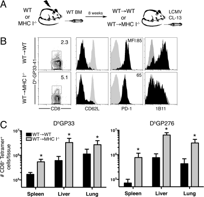
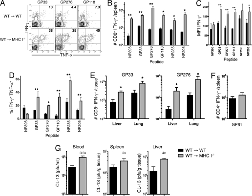
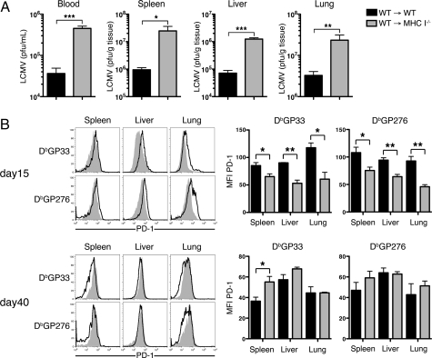
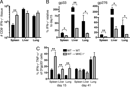
Many persistent viral infections induce dysfunctional T cell responses. Although a negative correlation exists between viral load and T cell responses during chronic infection, it is not known whether high antigen levels are the cause or just the consequence of T cell exhaustion. Furthermore, it is unclear what role antigen presentation by bone-marrow (BM) derived versus infected parenchymal cells has on T cell exhaustion. To address these issues, we examined the influence of antigen presentation by different cell types on CD8(+) T cell responses during persistent infection of mice with lymphocytic choriomeningitis virus (LCMV) clone 13. We generated BM chimeric mice, in which non-BM derived cells were MHC class I deficient. Virus-specific CD8(+) T cells in lymphoid and nonlymphoid tissues were increased in both number and ability to produce cytokines in these mice soon after infection. However, viral clearance from infected MHC I(-/-) parenchyma was significantly impaired, despite increased populations of cytokine producing CTL. The CD8(+) T cell response was overwhelmed by sustained antigen persistence, becoming increasingly exhausted within 4-6 weeks. Thus, we find that (i) sustained antigen presentation directly drives T cell exhaustion during a chronic viral infection, (ii) CTL require direct antigen-MHC interactions to clear virus-infected cells, and (iii) persistent interactions with antigen presented on both hematopoietic and nonhematopoietic cells negatively impacts virus-specific T cell responses during chronic infection.


Conflict of interest statement

The authors declare no conflict of interest.

Figures

Fig. 1.
Fig. 1.
Virus-specific CD8+ T cell responses in MHC I−/− BM chimeras. (A) MHC I−/− or wild-type (WT) mice were reconstituted with WT BM (WT → MHC Ι−/− or WT → WT) and infected with LCMV CL-13. (B) DbGP33-specific CD8+ T cells were analyzed in the spleen 15 days later. Expression of the indicated markers on Tetramer-specific CD8+ T cells (black histograms). Control staining of naïve CD8+ T cells from the mice is shown (gray histograms). (C) Quantitation of DbGP33- and DbGP276-specific T cells in the spleen, liver and lung 15 days after infection. *, P < 0.05; error bars represent SEM. One representative experiment of 3 is shown.
Fig. 2.
Fig. 2.
Enhanced cytokine production by virus-specific CD8+ T cells in MHC I−/− BM chimeras. (A) IFN-γ and TNF-α production by GP33-, GP276-, and GP118-specific CD8+ T cells from spleens of chimeric mice 15 days after LCMV CL-13 infection. Numbers represent the percentage of IFN-γ+ CD8+ T cells that are TNF-α+. (B) The number of splenic CD8+ T cells producing IFN-γ upon stimulation with either of 6 LCMV peptides as indicated. (C) Mean fluorescence intensity (MFI) of IFN-γ expression in the 6 antigen-specific CD8+ T cell populations after CL-13 infection. (D) TNF-α production by IFN-γ+ CD8+ T cells upon stimulation with the indicated peptides. (E) The number of gp33- or gp276-specific CD8+ T cells from the liver or lung producing IFN-γ upon stimulation. (F) Equivalent GP61-specific CD4+ T cell responses in the spleen after infection, as measured by production of IFN-γ by antigen-specific cells. (G) Viral titers in the blood, spleen and liver in chimeric mice 15 days after LCMV CL-13 infection. Numbers above WT → MHC I−/− bars represent fold increase in viral titer over that in WT → WT mice. *, P = <0.05; **, P = <0.01; error bars represent SEM. One representative experiment of 2–3 is shown.
Fig. 3.
Fig. 3.
Reduced viral clearance in mice lacking MHC I on nonhematopoietic cells. (A) Viral titers were measured in the serum, spleen, liver and lungs 30 days after LCMV CL-13 infection. (B) Changes in PD-1 expression on antigen-specific CD8+ T cells 2 and 6 weeks after infection. Representative expression of PD-1 on DbGP33- and DbGP276-specific CD8+ T cells in the indicated tissues from WT → WT mice (open histograms) and WT → MHC I−/− mice (filled histograms) is shown at Left. Graphs depict the mean fluorescence intensity (MFI) of PD-1 expression on the tetramer positive cells. *, P = <0.05; **, P = <0.01; error bars represent SEM. All histograms are representative of 2–3 independent experiments and summarized results are pooled from 2 experiments (n = 4–10 mice per group).
Fig. 4.
Fig. 4.
Persistent high levels of antigen drive functional exhaustion of T cells. (A) Total number of IFN-γ producing CD8+ T cells responding to 6 LCMV epitopes (NP396, GP33, GP276, GP118, NP235, and NP205) in the spleen, liver and lungs 41 days after CL-13 infection. (B) The frequency of CD8+ T cells producing IFN-γ after GP33 or GP276 peptide stimulation at day 41 postinfection, relative to that at day 15. (C) The proportion of DbGP33-specific CD8+ T cells producing IFN-γ and TNF-α in the indicated tissues, 15 or 41 days after CL-13 infection. *, P < 0.05; **, P < 0.01; error bars represent SEM. Data are representative of 2 independent experiments of 4–10 mice per group.

References

    1. Goepfert PA, et al. A significant number of human immunodeficiency virus epitope-specific cytotoxic T lymphocytes detected by tetramer binding do not produce gamma interferon. J Virol. 2000;74:10249–10255. - PMC - PubMed
    1. Gruener NH, et al. Sustained dysfunction of antiviral CD8+ T lymphocytes after infection with hepatitis C virus. J Virol. 2001;75:5550–5558. - PMC - PubMed
    1. Klenerman P, Hill A. T cells and viral persistence: Lessons from diverse infections. Nat Immunol. 2005;6:873–879. - PubMed
    1. Wherry EJ, Blattman JN, Murali-Krishna K, van der Most R, Ahmed R. Viral persistence alters CD8 T-cell immunodominance and tissue distribution and results in distinct stages of functional impairment. J Virol. 2003;77:4911–4927. - PMC - PubMed
    1. Zajac AJ, et al. Viral immune evasion due to persistence of activated T cells without effector function. J Exp Med. 1998;188:2205–2213. - PMC - PubMed

Publication types

MeSH terms