. 2009 Aug;19(8):899-909.
doi: 10.1093/glycob/cwp065. Epub 2009 May 11.

Targeted glycoproteomic identification of cancer cell glycosylation

Affiliations

Targeted glycoproteomic identification of cancer cell glycosylation

Alex S Powlesland et al. Glycobiology. 2009 Aug.

Abstract

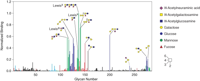
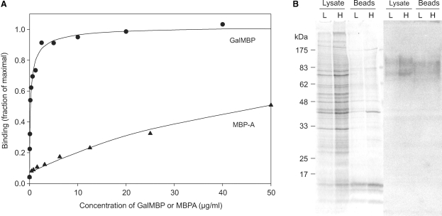
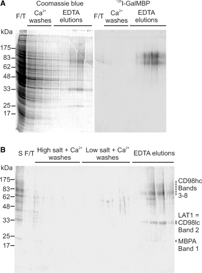
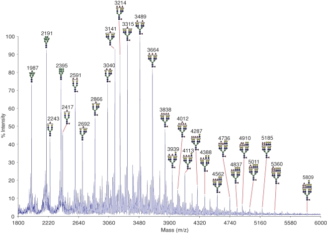
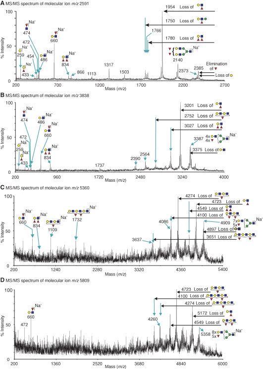
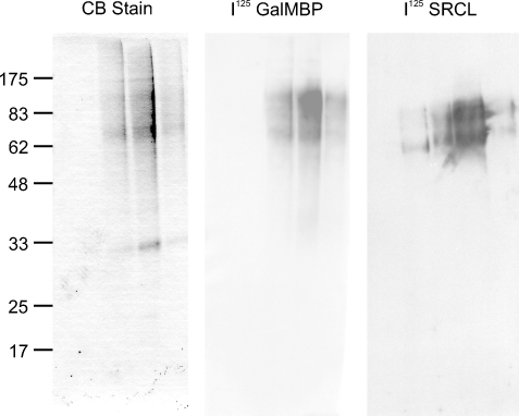
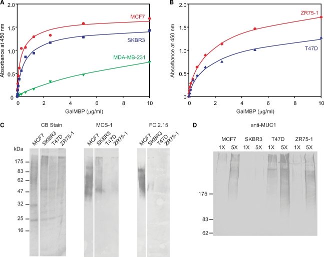
GalMBP is a fragment of serum mannose-binding protein that has been modified to create a probe for galactose-containing ligands. Glycan array screening demonstrated that the carbohydrate-recognition domain of GalMBP selectively binds common groups of tumor-associated glycans, including Lewis-type structures and T antigen, suggesting that engineered glycan-binding proteins such as GalMBP represent novel tools for the characterization of glycoproteins bearing tumor-associated glycans. Blotting of cell extracts and membranes from MCF7 breast cancer cells with radiolabeled GalMBP was used to demonstrate that it binds to a selected set of high molecular weight glycoproteins that could be purified from MCF7 cells on an affinity column constructed with GalMBP. Proteomic and glycomic analysis of these glycoproteins by mass spectrometry showed that they are forms of CD98hc that bear glycans displaying heavily fucosylated termini, including Lewis(x) and Lewis(y) structures. The pool of ligands was found to include the target ligands for anti-CD15 antibodies, which are commonly used to detect Lewis(x) antigen on tumors, and for the endothelial scavenger receptor C-type lectin, which may be involved in tumor metastasis through interactions with this antigen. A survey of additional breast cancer cell lines reveals that there is wide variation in the types of glycosylation that lead to binding of GalMBP. Higher levels of binding are associated either with the presence of outer-arm fucosylated structures carried on a variety of different cell surface glycoproteins or with the presence of high levels of the mucin MUC1 bearing T antigen.


Figures

Fig. 1
Fig. 1
Screening of a glycan array with GalMBP. Immobilized glycans were probed with fluorescein-labeled GalMBP. Glycans bearing terminal galactose are highlighted in red, except those that also contain fucose, which are colored green, and those linked via a β1,3 linkage to GalNAc, which are colored dark blue. Glycans bearing terminal GalNAc are indicated in light blue. The level of fluorescence was normalized to glycan 138. A full list of glycans present on the array is included in supplementary Table I.
Fig. 2
Fig. 2
GalMBP binding to MCF7 cells and glycoproteins. (A) Binding of GalMBP or wild-type MBP-A to MCF7 cells was detected using a rabbit polyclonal antibody to MBP-A, followed by a goat anti-rabbit secondary antibody conjugated to horseradish peroxidase. Experimental data (symbols) are shown together with curves fitted to the data using a direct binding equation. The concentration of protein required for 50% of maximal binding was approximately 0.1 μg/mL for GalMBP and 20 μg/mL for wild-type MBP-A. (B) Total cell lysates and biotinylated cell-surface proteins purified on avidin-conjugated beads were analyzed by SDS–polyacrylamide gel electrophoresis followed by Coomassie blue staining or blotting onto nitrocellulose followed by probing with 125I-GalMBP. The experiment was performed with low (L) and high (H) densities of MCF7 cells.
Fig. 3
Fig. 3
Purification of GalMBP ligands from MCF7 cell lysates by affinity chromatography on immobilized GalMBP. (A) Solubilized MCF7 cells were passed over a column containing immobilized GalMBP, which was washed in the presence of calcium and eluted with EDTA. Fractions were analyzed by SDS–polyacrylamide gel electrophoresis followed by staining with Coomassie blue or blotting onto nitrocellulose followed by probing with 125I-GalMBP. (B) GalMBP ligands were repurified on the GalMBP column and analyzed by SDS–polyacrylamide gel electrophoresis followed by staining with Coomassie blue. Bands from the elution fractions were excised and digested with trypsin. Proteins identified by mass spectrometry in each band are indicated.
Fig. 4
Fig. 4
Mass spectrometric analysis of N-glycans from CD98hc. Glycans released from purified GalMBP ligands were permethylated and subjected to MALDI MS profiling. All labeled molecular ions corresponding to complex-type glycans were subjected to MS/MS analysis. The assignment for each ion represents the most likely structure based on the precise fit between the calculated composition and the m/z value, taking into account the biosynthetic pathways of N-glycosylation in addition to the MS and MS/MS data. Values (m/z) listed correspond to 12C throughout. Satellite peaks surrounding major signals are either artifacts of the permethylation process or are minor glycans with one or more sialylated antennae. The symbols are defined in Figure 1.
Fig. 5
Fig. 5
MALDI-TOF/TOF spectra of permethylated N-glycans from CD98hc. Assignments of the fragment ions are shown either as loss of glycan moieties from the molecular ion (see annotations on horizontal arrows) or as antenna fragments which are B ions for cleavage at HexNAc and C ions for cleavage at hexoses (Domon and Costello 1988). Assigned masses correspond to the 12C isotope. The annotated ions not assigned in the schematics correspond to double or triple cleavages. Symbols are defined in Figure 1. (A) Fragmentation of the m/z 2591 molecular ion (Fuc3Hex5HexNAc4) produced ions at m/z 660 and 1954, corresponding to cleaving off of terminal Fuc-(Hex-HexNAc). The presence of terminal Lewisx is demonstrated by the diagnostic ion at m/z 2385, resulting from elimination of fucose attached at the 3 position of GlcNAc. (B) Fragments from the molecular ion at m/z 3838 (Fuc5Hex7HexNAc6) include ions at m/z 660, corresponding to Lewisx/a, m/z 834, corresponding to Lewisy/b, and m/z 474 corresponding to core fucosylation. (C) Dominant fragments from the ion at m/z 5360 (Fuc6Hex10HexNAc9), at m/z 1109, and at m/z 4274 indicate the presence of a two-repeat poly N-acetyllactosamine bearing one fucose residue. (D) Fragmentation of the ion at m/z 5809 (Fuc6Hex11HexNAc10) reveals the presence of three-repeat structures bearing one or two fucose residues at m/z 4100 and 4274.
Fig. 6
Fig. 6
Demonstration of GalMBP ligands from MCF7 cells binding to the scavenger receptor C-type lectin. Purified MCF7 ligands were analyzed by SDS–polyacrylamide gel electrophoresis. Parallel lanes were stained with Coomassie blue or blotted onto nitrocellulose membranes and probed with 125I-GalMBP or 125I-scavenger receptor C-type lectin (SRCL).
Fig. 7
Fig. 7
Binding of GalMBP to a panel of breast cancer cell lines and purification of glycoprotein ligands. (A and B) Binding of GalMBP was assayed as described in Figure 2A. (C and D) Gel electrophoresis of GalMBP ligands from multiple tumor cell lines purified by affinity chromatography on immobilized GalMBP. (C) Gels (17.5%) were stained with Coomassie blue or blotted onto nitrocellulose and probed with monoclonal anti-Lewisx antibodies followed by the peroxidase-conjugated goat anti-mouse secondary antibody. (D) Gels (5%) were blotted on nitrocellulose and probed with the monoclonal anti-MUC1 antibody followed by the peroxidase-conjugated goat anti-mouse secondary antibody.

References

    1. Abu-Qarn M, Yurist-Doutsch S, Giordano A, Trauner A, Morris HR, Hitchen P, Medalia O, Dell A, Eichler J. Haloferax volcanii AglB and AglD are involved in N-glycosylation of the S-layer glycoprotein and proper assembly of the surface layer. J Mol Biol. 2007;374:1224–1236. - PubMed
    1. Bray J, Lemieux RU, McPherson TA. Use of a synthetic hapten in the demonstration of the Thomsen-Friedenreich (T) antigen on neuraminidase-treated human red blood cells and lymphocytes. J Immunol. 1981;126:1966–1969. - PubMed
    1. Burchell JM, Mungul A, Taylor-Papadimitiriou J. O-Linked glycosylation in the mammary gland: Changes that occur during malignancy. J Mammary Gland Biol Neoplasia. 2001;6:355–364. - PubMed
    1. Burnette WN. “Western blotting”: Electrophoretic transfer of proteins from sodium dodecyl sulfate-polyacrylamide gels to unmodified nitrocellulose and radiographic detection with antibody and radioiodinated protein A. Anal Biochem. 1981;112:195–203. - PubMed
    1. Coombs PJ, Graham SA, Drickamer K, Taylor ME. Selective binding of the scavenger receptor C-type lectin to Lewisx trisaccharide and related glycan ligands. J Biol Chem. 2005;280:22993–22999. - PubMed

Publication types