. 2009;17(3):621-9.
doi: 10.3233/JAD-2009-1079.

Altered cerebral hemodynamics in early Alzheimer disease: a pilot study using transcranial Doppler

Affiliations

Altered cerebral hemodynamics in early Alzheimer disease: a pilot study using transcranial Doppler

Jurgen A H R Claassen et al. J Alzheimers Dis. 2009.

Abstract

Cerebrovascular disease may contribute to the development and progression of Alzheimer's disease (AD). This study investigated whether impairments in cerebral hemodynamics can be detected in early-stage AD. Nine patients with mild AD and eight cognitively normal controls matched for age underwent brain magnetic resonance imaging and neuropsychological evaluation, followed by assessment of steady-state cerebral blood flow velocity (CBFV, transcranial Doppler), blood pressure (BP, Finapres), and cerebrovascular resistance index (BP/CBFV). Cerebral hemodynamics were quantified using spectral and transfer function analysis of BP and CBFV in rest, during standing up after squat, and during repeated squat-stand maneuvers. Compared to controls, AD patients had lower CBFV and higher cerebrovascular resistance index, unexplained by brain atrophy. Low-frequency variability of BP was enhanced, suggesting impaired arterial baroreflex function. However, CBFV variability was reduced despite enhanced BP variability, and dynamic cerebral autoregulation was not impaired. In conclusion, despite a distinct pattern of altered cerebral hemodynamics, AD patients may have normal autoregulation. However, the challenges for autoregulation in AD are higher, as our data show enhanced BP fluctuations. Increased cerebral vasoconstriction or reduced vasomotion also may attenuate CBFV variability.


Figures

Fig. 1
Fig. 1
Spontaneous and induced oscillations in blood pressure and cerebral blood flow velocity. Example from two representative subjects, one with AD and one normal control, demonstrating spontaneous oscillations in blood pressure and cerebral blood flow-velocity over a period of 5 min (panel A). Panel B through D depict the effect of repeated squat-stand maneuvers at different intervals on these oscillations. Recording periods are 5, 4 and 3 min respectively. Panel B shows how repeated squat-stand intervals at 20 s lead to oscillations at 0.025 Hz. Panel C: 10 s intervals with oscillations at 0.05 Hz. Panel D: 5 s intervals with oscillations at 0.1 Hz.
Fig. 2
Fig. 2
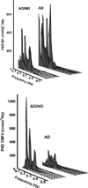
Spectral density plots of oscillations in blood pressure and cerebral blood flow velocity. Individual power spectral density plots for aged controls and AD patients, for blood pressure (upper pane), and cerebral blood flow-velocity (lower pane). Spectral power is a measure indicating the strength of oscillations at the specified frequency. The Y-axis shows the magnitude of the observed oscillations, and the X-axis displays the frequency of these oscillations. Note the much stronger oscillations in blood pressure in the very low frequency range (0.02–0.07 Hz) in AD patients, with reduced oscillations in cerebral blood flow-velocity.
Fig. 3
Fig. 3
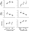
Correlation between spectral power of blood pressure (PSBP) and cerebral blood flow velocity (PSCBFV). A: spontaneous oscillations during rest. B: oscillations induced by repeated squat-stand maneuvers at 0.025, 0.05 and 0.1 Hz. For each individual, PSBP is plotted against PSCBFV. ■: AD patients. ●: normal controls. Filled symbols: very low frequency (0.02–0.07 Hz, A) and 0.025 and 0.05 Hz maneuvers (B), half-filled symbols: low frequency (0.07–0.2 Hz, A) and 0.1 Hz maneuvers (B), open symbols: high frequency (0.2–0.35 Hz, A). The correlation between PSBP and PSCBFV (p < 0.05) is less strong in AD patients than in controls. In addition, the slope of the regression lines differs between patients and controls (A: 1.18 vs. 0.23, B: 0.39 vs. 0.11, p = 0.01), indicating lower PSCBFV relative to PSBP in AD patients.
Fig. 4
Fig. 4
Transfer function analysis of spontaneous and induced oscillations in blood pressure and cerebral blood flow velocity. Group-averaged data showing results of transfer function analysis of spontaneous oscillations (left) in blood pressure (BP) and cerebral blood flow-velocity (CBFV) as well as of oscillations induced by repeated squat-stand maneuvers (right). ■: AD patients.●: normal controls. *: p < 0.05; ** p < 0.01. N = 9 (AD patients) resp. 8 (controls) for spontaneous oscillations and maneuvers at 0.05 and 0.1 Hz; n = 4 for maneuvers at 0.025 Hz.

References

    1. Suter OC, Sunthorn T, Kraftsik R, Straubel J, Darekar P, Khalili K, Miklossy J. Cerebral hypoperfusion generates cortical watershed microinfarcts in Alzheimer disease. Stroke. 2002;33:1986–1992. - PubMed
    1. Aliev G, Smith MA, Obrenovich ME, de la Torre JC, Perry G. Role of vascular hypoperfusion-induced oxidative stress and mitochondria failure in the pathogenesis of Alzheimer disease. Neurotox Res. 2003;5:491–504. - PubMed
    1. Park L, Anrather J, Forster C, Kazama K, Carlson GA, Iadecola C. Abeta-induced vascular oxidative stress and attenuation of functional hyperemia in mouse somatosensory cortex. J Cereb Blood Flow Metab. 2004;24:334–342. - PubMed
    1. Wu Z, Guo H, Chow N, Sallstrom J, Bell RD, Deane R, Brooks AI, Kanagala S, Rubio A, Sagare A, Liu D, Li F, Armstrong D, Gasiewicz T, Zidovetzki R, Song X, Hofman F, Zlokovic BV. Role of the MEOX2 homeobox gene in neurovascular dysfunction in Alzheimer disease. Nat Med. 2005;11:959–965. - PubMed
    1. Chow N, Bell RD, Deane R, Streb JW, Chen J, Brooks A, Van NW, Miano JM, Zlokovic BV. Serum response factor and myocardin mediate arterial hypercontractility and cerebral blood flow dysregulation in Alzheimer’s phenotype. Proc Natl Acad Sci U S A. 2007;104:823–828. - PMC - PubMed

Publication types