Review
. 2009 Dec;14(4):213-24.
doi: 10.1007/s10741-009-9146-x.

Abnormalities of calcium metabolism and myocardial contractility depression in the failing heart

Affiliations
Review

Abnormalities of calcium metabolism and myocardial contractility depression in the failing heart

Stephan E Lehnart et al. Heart Fail Rev. 2009 Dec.

Abstract

Heart failure (HF) is characterized by molecular and cellular defects which jointly contribute to decreased cardiac pump function. During the development of the initial cardiac damage which leads to HF, adaptive responses activate physiological countermeasures to overcome depressed cardiac function and to maintain blood supply to vital organs in demand of nutrients. However, during the chronic course of most HF syndromes, these compensatory mechanisms are sustained beyond months and contribute to progressive maladaptive remodeling of the heart which is associated with a worse outcome. Of pathophysiological significance are mechanisms which directly control cardiac contractile function including ion- and receptor-mediated intracellular signaling pathways. Importantly, signaling cascades of stress adaptation such as intracellular calcium (Ca(2+)) and 3'-5'-cyclic adenosine monophosphate (cAMP) become dysregulated in HF directly contributing to adverse cardiac remodeling and depression of systolic and diastolic function. Here, we provide an update about Ca(2+) and cAMP dependent signaling changes in HF, how these changes affect cardiac function, and novel therapeutic strategies which directly address the signaling defects.


Figures
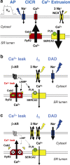
Fig. 1
Fig. 1
a Physiology of excitation-contraction (EC) coupling. An incoming action potential (AP) rapidly depolarizes the cell membrane potential (V m) in phase 0 through opening of voltage-dependent Na+ channels (NaV1.5). Subsequently, EC coupling is initiated through the opening of voltage-dependent L-type Ca2+ channels (CaV1.2) and the plasma membrane Ca2+ influx current (I Ca). I Ca activates ryanodine receptors (RyR2s) and intracellular Ca2+ release from sarcoplasmic reticulum (SR) stores, also known as Ca2+ induced Ca2+ release (CICR). CICR is followed by extrusion of Ca2+ from the cytosol into the SR by Ca2+ pumps (SERCA2) and to the extracellular compartment by the Na+/Ca2+ exchanger (NCX) operating in its forward mode (3 Na+ in for each Ca2+ out), which creates an electrogenic inward current. SR Ca2+ leak is inhibited by the calstabin2 (Cab2) subunit which stabilizes the RyR2 closed state. The SERCA2 pump rate is inhibited by the phospholamban (PLN) subunit in its dephosphorylated state. Ca2+ release and uptake occur cyclically during each heart beat and represent 60–90% of Ca2+ signaling during EC coupling depending on the species studied. b EC coupling abnormalities in CPVT. RyR2 missense mutations significantly increase the propensity for intracellular Ca2+ leak in resting cardiomyocytes (during diastole) with filled SR Ca2+ stores. Stimulation of β-adrenergic receptors (β-ARs) during stress adaptation results in RyR2 and PLN phosphorylation by PKA (indicated by ⊗) which increases SR Ca2+ release and uptake, respectively. However, RyR2 mutations (as indicated by green star) in the PKA phosphorylated Ca2+ release channel lead to partial calstabin2 depletion, a significant gain-of-function defect of RyR2, and intracellular Ca2+ leak. RyR2 Ca2+ leak activates depolarizing transient inward currents (I TI) supposedly through abnormal forward mode NCX activity. If I TI currents reach a critical threshold of membrane potential instability in phase 4 of the cardiac AP, Na+ channels are activated leading to delayed after depolarizations (DADs) which underly triggered activity. c EC coupling abnormalities in HF. HF is a chronic hyperadrenergic state which results in downregulation of β-AR signaling and reduced intracellular cAMP synthesis. However, maintained hyperadrenergic stimulation of β-ARs during HF results in chronic RyR2 PKA hyperphosphorylation (indicated by large ⊗), depletion of the stabilizing calstabin2 subunits as well as other components of the channel complex including phosphodiesterase 4D3 (PDE4D3). PDE4D3 depletion causes a chronically reduced cAMP hydrolysis in the channel complex and contributes to RyR2 PKA hyperphosphorylation induced intracellular Ca2+ leak. On the other hand, PLN is chronically PKA hypophosphorylated (indicated by small ⊗) creating constitutively inhibited state of SERCA2 and reduced SR Ca2+ uptake. Additionally, NCX expression is significantly increased leading to abnormally increased Ca2+ extrusion to the extracellular side and depletion of intracellular Ca2+ stores. Despite Ca2+ store depletion, DADs and triggered activity are frequent in HF possibly due to increased SR Ca2+ leak and proarrhythmogenic inward NCX and late I Na,L currents
Fig. 2
Fig. 2
Representative traces of aequorin-based Ca2+ signals and corresponding isometric forces from human nonfailing (top) and failing (bottom) myocardial muscle preparations. Upper panel: Nonfailing myocardium shows post-rest potentiation of the intracellular Ca2+ transient and force development which increases from 10 to 120 s rest period. Lower panel: Failing myocardium shows depressed post-rest intracellular Ca2+ transient and force development after 120 s rest. Steady-state pre-rest signals are shown on the left of each trace; first and second post-rest signals are shown afterwards; post-rest signals represent 10 s (left) and 120 s (right), dimensions as indicated. Reproduced with permission from the Journal of Clinical Investigation (Pieske et al. [80])

References

    1. Bristow MR, Ginsburg R, Umans V, Fowler M, Minobe W, Rasmussen R, Zera P, Menlove R, Shah P, Jamieson S, et al. Beta 1- and beta 2-adrenergic-receptor subpopulations in nonfailing and failing human ventricular myocardium: coupling of both receptor subtypes to muscle contraction and selective beta 1-receptor down-regulation in heart failure. Circ Res. 1986;59:297–309. - PubMed
    1. Cohn JN, Levine TB, Olivari MT, Garberg V, Lura D, Francis GS, Simon AB, Rector T. Plasma norepinephrine as a guide to prognosis in patients with chronic congestive heart failure. N Engl J Med. 1984;311:819–823. - PubMed
    1. Feldman MD, Copelas L, Gwathmey JK, Phillips P, Warren SE, Schoen FJ, Grossman W, Morgan JP. Deficient production of cyclic AMP: pharmacologic evidence of an important cause of contractile dysfunction in patients with end-stage heart failure. Circulation. 1987;75:331–339. - PubMed
    1. Foo RS, Mani K, Kitsis RN. Death begets failure in the heart. J Clin Invest. 2005;115:565–571. - PMC - PubMed
    1. Wencker D, Chandra M, Nguyen K, Miao W, Garantziotis S, Factor SM, Shirani J, Armstrong RC, Kitsis RN. A mechanistic role for cardiac myocyte apoptosis in heart failure. J Clin Invest. 2003;111:1497–1504. - PMC - PubMed

Publication types

MeSH terms

Substances