Deformation of nasal septal cartilage during mastication
- PMID: 19434723
- PMCID: PMC2786896
- DOI: 10.1002/jmor.10750
Deformation of nasal septal cartilage during mastication
Abstract
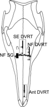
The cartilaginous nasal septum plays a major role in structural integrity and growth of the face, but its internal location has made physiologic study difficult. By surgically implanting transducers in 10 miniature pigs (Sus scrofa), we recorded in vivo strains generated in the nasal septum during mastication and masseter stimulation. The goals were (1) to determine whether the cartilage should be considered as a vertical strut supporting the nasal cavity and preventing its collapse, or as a damper of stresses generated during mastication and (2) to shed light on the overall pattern of snout deformation during mastication. Strains were recorded simultaneously at the septo-ethmoid junction and nasofrontal suture during mastication. A third location in the anterior part of the cartilage was added during masseter stimulation and manipulation. Contraction of jaw closing muscles during mastication was accompanied by anteroposterior compressive strains (around -1,000 muepsilon) in the septo-ethmoid junction. Both the orientation and the magnitude of the strain suggest that the septum does not act as a vertical strut but may act in absorbing loads generated during mastication. The results from masseter stimulation and manipulation further suggest that the masticatory strain pattern arises from a combination of dorsal bending and/or shearing and anteroposterior compression of the snout.
2009 Wiley-Liss, Inc.
Figures


Similar articles
-
Compressive and tensile mechanical properties of the porcine nasal septum.J Biomech. 2014 Jan 3;47(1):154-61. doi: 10.1016/j.jbiomech.2013.09.026. Epub 2013 Oct 8. J Biomech. 2014. PMID: 24268797 Free PMC article.
-
Susan Herring profile. Getting inside your head.Science. 2004 Oct 29;306(5697):804-5. doi: 10.1126/science.306.5697.804. Science. 2004. PMID: 15514132 No abstract available.
-
Mastication and the postorbital ligament: dynamic strain in soft tissues.Integr Comp Biol. 2011 Aug;51(2):297-306. doi: 10.1093/icb/icr023. Epub 2011 May 18. Integr Comp Biol. 2011. PMID: 21593142 Free PMC article.
-
Jaw muscles and the skull in mammals: the biomechanics of mastication.Comp Biochem Physiol A Mol Integr Physiol. 2001 Dec;131(1):207-19. doi: 10.1016/s1095-6433(01)00472-x. Comp Biochem Physiol A Mol Integr Physiol. 2001. PMID: 11733178 Review.
-
Anatomy and Physiology of the Nasal Valves.Otolaryngol Clin North Am. 2025 Apr;58(2):189-203. doi: 10.1016/j.otc.2024.09.001. Epub 2024 Oct 18. Otolaryngol Clin North Am. 2025. PMID: 39426874 Review.
Cited by
-
Compressive and tensile mechanical properties of the porcine nasal septum.J Biomech. 2014 Jan 3;47(1):154-61. doi: 10.1016/j.jbiomech.2013.09.026. Epub 2013 Oct 8. J Biomech. 2014. PMID: 24268797 Free PMC article.
-
Properties of the Nasal Cartilage, from Development to Adulthood: A Scoping Review.Cartilage. 2022 Jan-Mar;13(1):19476035221087696. doi: 10.1177/19476035221087696. Cartilage. 2022. PMID: 35345900 Free PMC article.
-
The biomechanical role of the chondrocranium and sutures in a lizard cranium.J R Soc Interface. 2017 Dec;14(137):20170637. doi: 10.1098/rsif.2017.0637. J R Soc Interface. 2017. PMID: 29263126 Free PMC article.
-
Evaluation of human nasal cartilage nonlinear and rate dependent mechanical properties.J Biomech. 2020 Feb 13;100:109549. doi: 10.1016/j.jbiomech.2019.109549. Epub 2019 Nov 29. J Biomech. 2020. PMID: 31926590 Free PMC article.
-
Structure and function of the septum nasi and the underlying tension chord in crocodylians.J Anat. 2016 Jan;228(1):113-24. doi: 10.1111/joa.12404. Epub 2015 Nov 10. J Anat. 2016. PMID: 26552989 Free PMC article.
References
-
- Badoux DM. Framed structures in the mammalian skull. Acta Morphol Neerl Scand. 1966;6:239–250. - PubMed
-
- Badoux DM. Cremona diagrams of framed structures in skull of Canis familiaris and Sus scrofa scrofa. Koninkl Nederl Akad van Wetenschappen C. 1968;71:229–244.
-
- Beynnon BD, Fleming BC. Anterior cruciate ligament strain in-vivo: A review of previous work. J Biomech. 1998;31:519–525. - PubMed
-
- Byl C, Puttlitz C, Byl N, Lotz J, Topp K. Strain in the median and ulnar nerves during upper-extremity positioning. J Hand Surg. 2002;27:1032–1040. - PubMed
-
- Cerulli G, Benoit DL, Lamontagne M, Caraffa A, Liti A. In vivo anterior cruciate ligament strain behaviour during a rapid deceleration movement: case report. Knee Surg Sports Traumatol Arthrosc. 2003;11:307–311. - PubMed
Publication types
MeSH terms
Grants and funding
LinkOut - more resources
Full Text Sources

