. 2009 Jul 14;48(27):6450-60.
doi: 10.1021/bi900174v.

A hotspot of inactivation: The A22S and V108M polymorphisms individually destabilize the active site structure of catechol O-methyltransferase

Affiliations

A hotspot of inactivation: The A22S and V108M polymorphisms individually destabilize the active site structure of catechol O-methyltransferase

Karen Rutherford et al. Biochemistry. .

Abstract

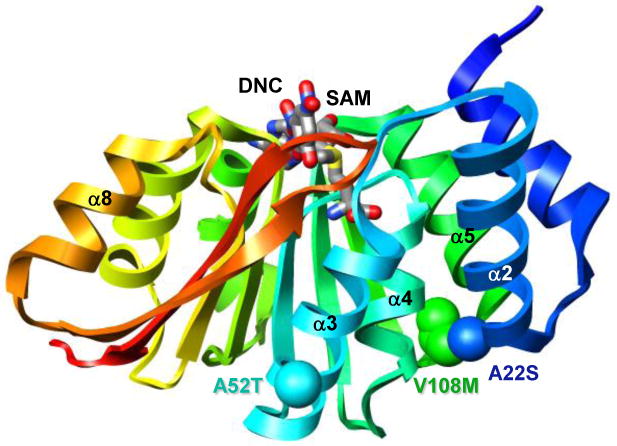
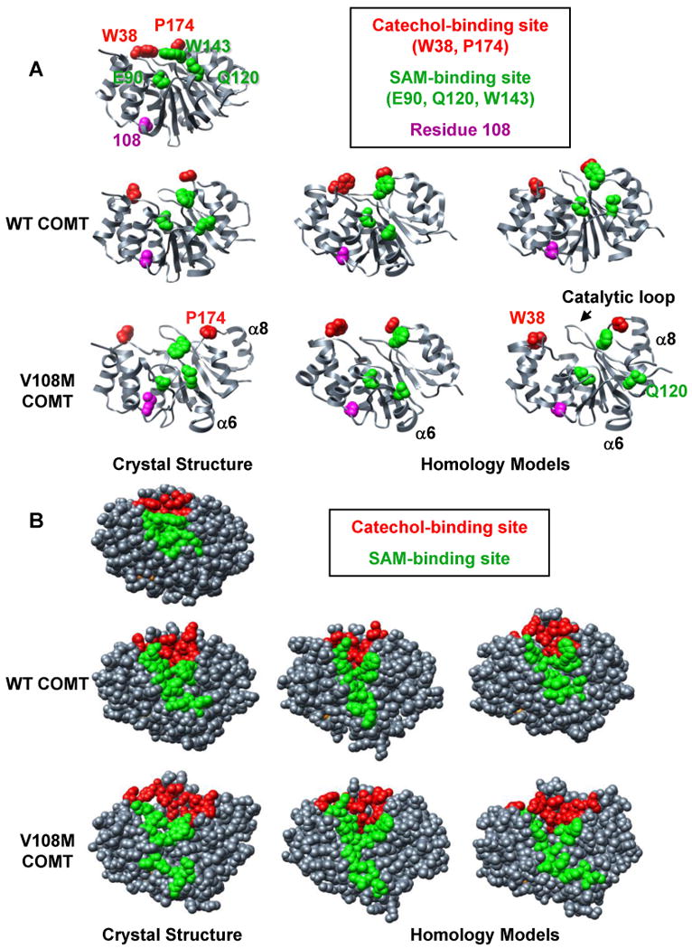
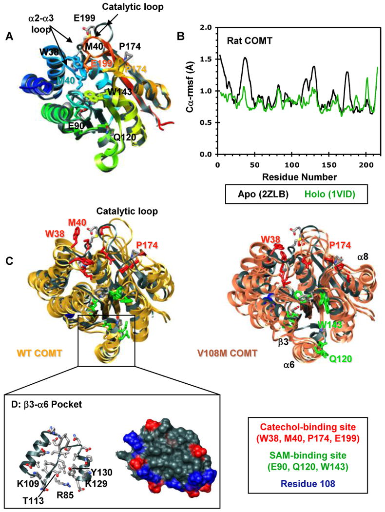
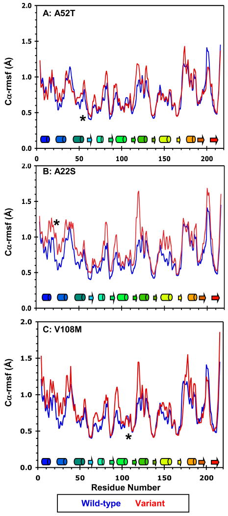
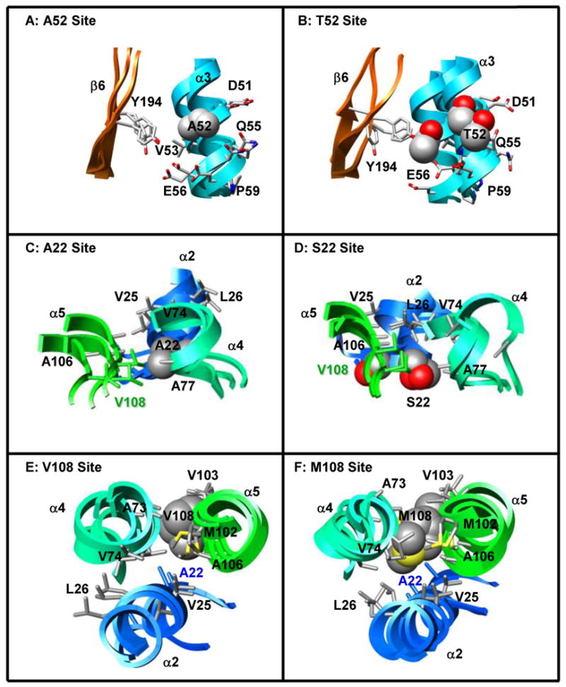
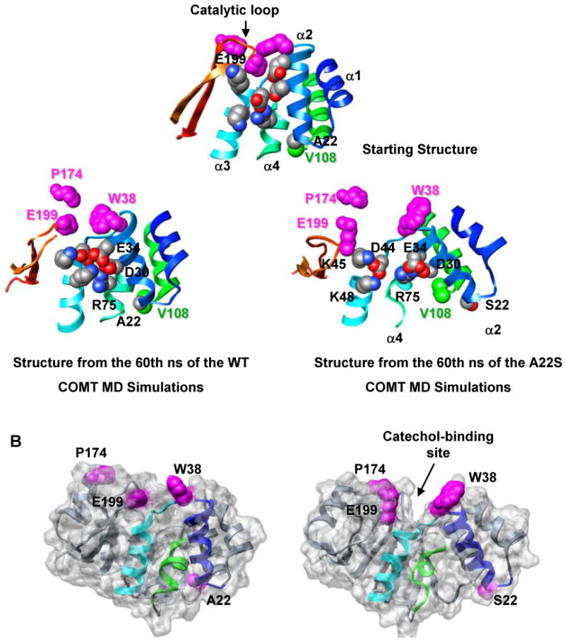
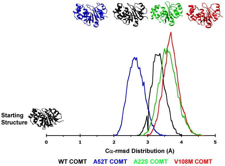
Human catechol O-methyltransferase (COMT) contains three common polymorphisms (A22S, A52T, and V108M), two of which (A22S and V108M) render the protein susceptible to deactivation by temperature or oxidation. We have performed multiple molecular dynamics simulations of the wild-type, A22S, A52T, and V108M COMT proteins to explore the structural consequences of these mutations. In total, we have amassed more than 1.4 micros of simulation time, representing the largest set of simulations detailing the effects of polymorphisms on a protein system to date. The A52T mutation had no significant effect on COMT structure in accord with experiment, thereby serving as a good negative control for the simulation set. Residues 22 (alpha2) and 108 (alpha5) interact with each other throughout the simulations and are located in a polymorphic hotspot approximately 20 A from the active site. Introduction of either the larger Ser (22) or Met (108) tightens this interaction, pulling alpha2 and alpha5 toward each other and away from the protein core. The V108M polymorphism rearranges active-site residues in alpha5, beta3, and alpha6, increasing the S-adenosylmethionine site solvent exposure. The A22S mutation reorients alpha2, moving critical catechol-binding residues away from the substrate-binding pocket. The A22S and V108M polymorphisms evolved independently in Northern European and Asian populations. While the decreased activities of both A22S and V108M COMT are associated with an increased risk for schizophrenia, the V108M-induced destabilization is also linked with improved cognitive function. These results suggest that polymorphisms within this hotspot may have evolved to regulate COMT activity and that heterozygosity for either mutation may be advantageous.


Figures

Figure 1
Figure 1
The A22S, A52T, and V108M COMT polymorphisms. Ribbon diagram of wild-type COMT (PDB entry 3BWM (54)) colored from blue (N-terminus) to red (C-terminus). S-adenosylmethionine (SAM) and 3,5-dinitrocatechol (DNC) are shown in stick representation and colored by atom type. Polymorphic residues 22, 52, and 108 are shown in space-filling representation and colored in blue, cyan and green, respectively.
Figure 2
Figure 2
Comparison of MD simulations performed at 37°C using crystal structures and homology models of human wild-type and V108M COMT. Snapshots of structures from the final ns of MD simulations of (left) the crystal structures of human wild-type (3BWM.PDB, (54)) and V108M (3BWY.PDB, (54)) COMT, and (center, right) the homology models of human COMT based on the crystal structure of rat COMT (1VID.PDB, (53)) performed with C-scale values of either 0.0 (center) or 0.4 (right) (see Materials and Methods). (A) The 108M COMT structure is prone to disruption at 37°C. Altered packing around residue M108 reorients helix α6, pulling Q120 away from the SAM-binding site. Helix α8 (and P174) pulls away from the protein core resulting in an expanded conformation. These motions occur during simulations of both the crystal structure and homology models of the V108M COMT protein, but are not observed during the simulations of wild-type COMT. SAM-binding residues (E90, Q120, W143), catechol-binding residues (W38, P174) and residue 108 are shown in space-filling representation and colored in green, red, and magenta, respectively. (B) The 108M COMT active site is more exposed to solvent and distorted at 37°C than that of the wild-type protein. Residues in the SAM- and catechol-binding sites are colored green and red, respectively.
Figure 3
Figure 3
Structural characterization of the COMT apoprotein. (A) Superposed ribbon diagrams of the rat COMT apoprotein (gray, 2ZLB.PDB (66)) and the rat (1VID.PDB (53)) and human (3BWM.PDB (54)) holoproteins bound with SAM and DNC and colored from blue (N-terminus) to red (C-terminus). The overall structures of the rat and human holoproteins are virtually identical. The rat apo and holoproteins differ mainly in the loop regions that define the catechol-binding site. The α2-α3 loop, β5-α8 loop, and the catalytic loop (residues 197-202) pull away from the protein core in the apoprotein exposing the SAM- and catechol-binding sites. This open configuration may facilitate substrate binding. Interestingly, the adenosine pocket of the apo and holoprotein SAM-binding sites are very similar showing only a slight displacement of key active site residues (E90, Q120, W143). Active site residues are shown in stick representation and colored/labeled in either gray (apoprotein) or to match the structure (holoprotein). (B) Cα-rmsf values (Å) per residue for the rat apoprotein (black, 2ZLB.PDB (66)) and holoprotein (green, 1VID.PDB (53)). (C) Structural overlay of the rat apoprotein crystal structure (gray, 2ZLB.PDB (66)) with structures from the final ns of three independent simulations of human WT (gold) and V108M (pink) COMT. The human WT and rat apoproteins behave similarly in that the structure of the adenosine pocket in the SAM-binding site (α5, α6, α7) is maintained, with some flexibility in the orientations of active site residues E90, Q120, and W143, while the α2-α3, β5-α8, and catalytic loops fluctuate greatly opening up the active-site core and moving W38, M40, P174 and E199 out of the substrate-binding pocket. In addition to these motions, both β3 and α6 pull away from the protein core during simulations of V108M COMT. SAM- and catechol-binding residues are shown in stick representation and colored in green and red, respectively. Residue 108 is shown in space-filling representation and colored blue. (D) Ribbon (left) and van der Waals surface (surface) representation of the β3-α6 pocket from human WT COMT. The V108M polymorphism alters the orientations of β3 and α6, possibly destabilizing the protein. Therefore, the β3-α6 pocket may serve as a target site for docking small molecules to stabilize the V108M COMT structure. Positively and negatively charged residues in the surface representation are colored in blue and red.
Figure 4
Figure 4
Mobility of the wild-type, A22S, A52T, and V108M COMT variants at 37°C. Cα-rmsf values (Å) per residue for the wild-type protein (blue) compared with those of the (A) A52T, (B) A22S, and (C) V108M COMT variants (all in red). Cα-rmsf values were calculated relative to the average structure over the last 10 ns of each simulation. Secondary structural elements are depicted as cylinders for α-helices and arrows for β-strands and are colored to match the structure in Figure 1. The polymorphic residues are marked by asterisks.
Figure 5
Figure 5
Effects of the A52T, A22S, and V108M COMT polymorphisms on local tertiary structure. (A, B) A52 (α3) forms mainchain hydrogen bonds with K48, I49, and E56 of α3, and sidechain contacts with residues in α3 (I49, V53, Q55, E56) and β6 (Y194). T52 maintains all of these contacts, while T52 OG1 forms additional hydrogen bonds with the backbone carbonyl groups of K48 and I49. (C, D) A22 is positioned in a loop between α1 and α2 where it interacts with residues in α2 (Q23, V25, L26), α4 (V74, A77, R78), and α5 (A106, V108). The larger S22 forms an additional S22 OG – G107 O hydrogen bond enabling closer contact between S22 and residues in α5 (A106, G107, V108). This altered packing moves S22 away from residues V74 and R78 of α4, affecting the orientations of both α2 and α4 relative to their positions in the wild-type protein. (E, F) Residue 108 is located in a loop between α5 and β3. Both V108 and M108 contact residues in α2 (A22), α4 (A73, V74, R78), and α5 (V103, A106). The larger methionine forms additional contacts with V25 and L26 of α2 and M102 of α5, effectively moving helices α2 and α5 closer together. Structures from the final ns of three independent simulations are superposed in A-F and colored to match Figure 1. Sidechains of the polymorphic residues and their contacts are shown in space-filling and stick representations, respectively.
Figure 6
Figure 6
The A22S polymorphism disrupts the catechol-binding site. (A) The altered packing around residue S22 causes α2 and α5 to move towards each other while the last turn of α4 pulls away from the polymorphic site. This rearrangement results in the formation of new salt bridges between D30 (α2) – R75 (α4), E34 (α2) – R75 (α4), and E34 (α2) – K48 (α3). The altered packing around the polymorphic residue is propagated through α3 towards the protein's C-terminus leading to an overall expansion of the protein and the separation of key catechol-binding residues W38, P174, and E199. Residue 22, and residues involved in salt bridges are shown in space-filling representation and colored by atom type. V108 and catechol-binding residues are colored in green and magenta, respectively. Structures are colored as in Figure 1. (B) The rearrangement of α2 and α3 both increases the mobility of several residues in the α2-α3 loop including W38, a gate-keeping residue involved in catechol binding, and pulls these residues away from the protein's C-terminus. This movement opens a large cleft in the protein, which greatly increases the solvent exposure of the catechol-binding site, relative to the wild-type protein. Residue 22 and the catechol-binding residues (W38, P174, E199) are shown in space-filling representation and colored magenta. α2, α3, and α4 are colored as in Figure 1.
Figure 7
Figure 7
Total Cα–rmsd distributions of structures from MD simulations of wild-type, A22S, A52T, and V108M COMT. Structures from the WT COMT simulations fall into a narrow distribution centered at 3.3 Å Cα-rmsd from the starting structure. The 52T distribution is centered at 2.6 Å Cα-rmsd indicating that the A52T polymorphism somewhat stabilizes the protein structure. However, structures from the 52T COMT simulations fall into a broader distribution than those of the WT protein. Both the 22S and 108M COMT variants exist as ensembles of structures that are more distorted than those of the WT protein. The vertical scale is arbitrary.

References

    1. Axelrod J, Szara S. Enzymic conversion of metanephrine to epinephrine. Biochim Biophys Acta. 1958;30:188–189. - PubMed
    1. Mannisto PT, Kaakkola S. Catechol-O-methyltransferase (COMT): biochemistry, molecular biology, pharmacology, and clinical efficacy of the new selective COMT inhibitors. Pharmacol Rev. 1999;51:593–628. - PubMed
    1. Weinshilboum RM, Otterness DM, Szumlanski CL. Methylation pharmacogenetics: catechol O-methyltransferase, thiopurine methyltransferase, and histamine N-methyltransferase. Annu Rev Pharmacol Toxicol. 1999;39:19–52. - PubMed
    1. Tenhunen J, Salminen M, Jalanko A, Ukkonen S, Ulmanen I. Structure of the rat catechol-O-methyltransferase gene: separate promoters are used to produce mRNAs for soluble and membrane-bound forms of the enzyme. DNA Cell Biol. 1993;12:253–263. - PubMed
    1. Tenhunen J, Salminen M, Lundstrom K, Kiviluoto T, Savolainen R, Ulmanen I. Genomic organization of the human catechol O-methyltransferase gene and its expression from two distinct promoters. Eur J Biochem. 1994;223:1049–1059. - PubMed

Publication types

MeSH terms

Substances