. 2009 May 12:8:99.
doi: 10.1186/1475-2875-8-99.

PfeIK1, a eukaryotic initiation factor 2alpha kinase of the human malaria parasite Plasmodium falciparum, regulates stress-response to amino-acid starvation

Affiliations

PfeIK1, a eukaryotic initiation factor 2alpha kinase of the human malaria parasite Plasmodium falciparum, regulates stress-response to amino-acid starvation

Clare Fennell et al. Malar J. .

Abstract

Background: Post-transcriptional control of gene expression is suspected to play an important role in malaria parasites. In yeast and metazoans, part of the stress response is mediated through phosphorylation of eukaryotic translation initiation factor 2alpha (eIF2alpha), which results in the selective translation of mRNAs encoding stress-response proteins.

Methods: The impact of starvation on the phosphorylation state of PfeIF2alpha was examined. Bioinformatic methods were used to identify plasmodial eIF2alpha kinases. The activity of one of these, PfeIK1, was investigated using recombinant protein with non-physiological substrates and recombinant PfeIF2alpha. Reverse genetic techniques were used to disrupt the pfeik1 gene.

Results: The data demonstrate that the Plasmodium falciparum eIF2alpha orthologue is phosphorylated in response to starvation, and provide bioinformatic evidence for the presence of three eIF2alpha kinases in P. falciparum, only one of which (PfPK4) had been described previously. Evidence is provided that one of the novel eIF2alpha kinases, PfeIK1, is able to phosphorylate the P. falciparum eIF2alpha orthologue in vitro. PfeIK1 is not required for asexual or sexual development of the parasite, as shown by the ability of pfeik1- parasites to develop into sporozoites. However, eIF2alpha phosphorylation in response to starvation is abolished in pfeik1- asexual parasites

Conclusion: This study strongly suggests that a mechanism for versatile regulation of translation by several kinases with a similar catalytic domain but distinct regulatory domains, is conserved in P. falciparum.


Figures

Figure 1
Figure 1
The P. falciparum eIF2α orthologue is phosphorylated in response to amino-acid starvation. A: Alignment of PfeIF2α with orthologous sequences from T. gondii (Tg), human (Hs), rice (Os) and E. cuniculi (Ec). Sequences surrounding the conserved regulatory serine, (P. falciparum numbering: M48 – K108) are shown. Residues that are identical in all sequences are highlighted in black, residues that are identical or similar are marked in grey. The arrow indicates the serine that is the target of eIF2α kinases. Open arrow heads (∨) indicate residues involved in contacting the kinase domain, asterisks (*) indicate conserved residues that protect the phosphorylation site from the activity of other kinases. B. Western blot analysis of PfeIF2α phosphorylation. A 3D7 parasite culture synchronized to the late ring stage was equally partitioned into individual cultures. Growth of the parasites was continued up to 5 hours at 37°C in either complete RPMI medium (CM) or in RPMI lacking amino acids (-AA). CM was added back to one amino acid-deprived culture, and re-incubated for an additional 45 minutes. Total lysates from the parasites were prepared for SDS-PAGE, followed by immunoblotting with antibodies against phosphorylated eIF2α (anti-phospho eIF2α) and the endoplasmic reticulum (ER) marker, BiP (anti-BiP), which served as the loading control.
Figure 2
Figure 2
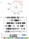
Bioinformatic analyses of P. falciparum eIF2α kinases. A: Phylogenetic tree showing clustering of PfeIF2α kinases with human eIF2α kinases. Sequences: PfeIK1: PF14_0114; PfeIK2: PFA0380w; PfPK4: PFF1370; PKR: GI:4506103; HRI: GI:6580979; PERK: GI:18203329; GCN2: GI:65287717; MEK1: GI:400274; IRAK1: GI:68800243; Aurora: GI:37926805; CDK2: GI:1942427; PfCK1: PF11_0377; PfNEK2: Pfe1290w; PfPK5: MAL13P1.279; PfPKA: PFI1685w; PFTKL3: PF13_0258; Pfb0815w: PfCDPK1; hCAMK1: GI:4502553; hPRKACA: GI:46909584; hCSNK1d: GI:20544145; hNEK7: GI:19424132; hSRC: GI:4885609. B. Alignment of the catalytic domains of PfeIK1, PbeIK1 and human GCN2. Identical residues in all three kinases are in black boxes, residues that are identical in two sequences of the three sequences, or that are similar are boxed in grey. The number of residues comprising the inserts between domains IV and V are marked between //-//. Asterisks (*) mark residues conserved among kinases in general, while open arrowheads (∨) indicate residues specifically conserved among eIF2α kinases. The downwards arrow marks the threonine residues that are targets for autophosphorylation in GCN2. PlasmoDB accession numbers: PfeIK1: PF14_0423, PbeIK1: PB000582.03.0 GenBank accession number: HsGCN2: GI:65287717. C: Schematic of the domain structures of PfeIK1, PbeIK1 and GCN2. Kinase domains (KD) are in grey, hatched regions represent the inserts (I) within the kinase domains and regions with no identified function are white. Additional characterized domains of GCN2 are as follows: red; N-terminal GCN1 binding domain (GB), green; pseudo-kinase domain (ΨKD), blue; histidyl-tRNA synthetase (HisRS), yellow; ribosome binding and dimerisation domain (RB/DD).
Figure 3
Figure 3
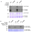
Kinase activity of PfeIK1. A: GST-PfeIK1 phosphorylates the exogenous substrates α-and β-casein. Kinase assays were performed using 10 μg α-casein (left 3 lanes) or β-casein (right 3 lanes), in the presence of 2 μg wild-type kinase catalytic domain (WT), catalytically inactive mutant (K458M) or no kinase (-). Upper panel: autoradiogram, lower panel: Coomassie blue-stained gel. B: GST-PfeIK1 autophosphorylates and can phosphorylate recombinant GST-PfeIF2α, but not the mutant GST-PfeIF2α-S59A. Kinase assays were performed using 2 μg wild-type PfeIK1 catalytic domain (WT), or catalytically inactive mutant (K458M), or no kinase (-), in the presence of 10 μg wild-type GST-PfeIF2α (left 3 lanes), targeted mutant GST-PfeIF2α-S59A (middle 3 lanes) or no substrate (right 2 lanes). The position of the substrate is highlighted by ovals.
Figure 4
Figure 4
Disruption of the pfeik1 gene. A. Strategy for gene disruption. The transfection plasmid contains a PCR fragment spanning positions 1467–2255 of the entire 4.8 kb pfeik1 coding sequence (as predicted on PlasmoDB). The fragment excludes two regions essential for catalytic activity, labelled 'ATP' (a glycine-rich region required for orientation of ATP) and 'E' (a glutamate residue required for structural stability of the enzyme). The positions of primers used for genotyping clones, and for nested PCR to genotype oocsyts are indicated by numbered arrows. B: PCR analysis. Genomic DNA isolated from pfeik1- clones C1 and C8, and from 3D7 wild-type parasites, was subjected to PCR using the indicated primers (see Figure 4A for primer locations). Lanes 1, 5, 9: primers 1 + 2 (diagnostic for the wild-type locus); lanes 2, 6, 10: primers 3 + 4 (diagnostic for the pCAM-BSD-PfeIK1 plasmid); lanes 3, 7, 11: primers 1 + 4 (diagnostic for 5' integration boundary); lanes 4,8,12: primers 3 + 2 (diagnostic for 3' integration boundary). M = co-migrating markers. C: Schematic of expected sizes on Southern blot analysis of wild-type 3D7 parasites and pfeik1- parasites. D: Southern blot analysis of the pfeik1 locus in wild-type 3D7 and pfeik1- clones C1 and C8. Genomic DNA was digested with HindIII, transferred to a Hybond membrane and probed with the pfeik1 fragment that was used as the insert in the pCAM-BSD-PfeIK1 plasmid. Positions of the bands corresponding to the wild-type locus (WT), 5' integration (5' int.), 3' integration (3' int.) and linearized plasmid (plasmid) are shown on the right. Sizes of co-migrating markers are indicated on the left.
Figure 5
Figure 5
Disruption of the pfeik1 gene does not affect asexual growth rate. Representative cycles of pfeik1-parasites and the parental 3D7 strain (dashed). Cycle points were semi-automatically collected fixed and stored at 4°C every 30 min over ~4 days. After permeabilization and RNAse treatment, the DNA content was analyzed by flow cytometry as previously described [42]. Mature schizonts (~16–32 N), red line; S-phase (~2–8 N), blue line; G1-phase (1N), black line. Percentage values as a function of time are shown; hpi: hours post-infection, referring to the mature schizont maxima as zero.
Figure 6
Figure 6
pfeIK1- parasites do not phosphorylate eIF2α in amino acid-limiting conditions. pfeik1- clones C1 and E6, as well as the 3D7 parent clone and a pfeik2- clone used as controls, were synchronized to the late ring stage and equally partitioned into individual cultures. Growth of the parasites was continued up to 5 hours at 37°C in either complete RPMI (CM) or in RPMI lacking amino acids (-AA). CM was added back to one amino acid-deprived culture, and re-incubated for an additional 45 minutes. Total lysates from the parasites were prepared for SDS-PAGE, followed by immunoblotting with antibodies against phosphorylated eIF2α (anti-phosho eIF2α). Antibodies against the endoplasmic reticulum marker BiP (anti-BiP) served as the loading control.
Figure 7
Figure 7
Analysis of the parasite genotypes in mosquito infections. Genomic DNA extracted from a wild-type 3D7-infected mosquito and from two mosquitoes infected with clone C8 was analysed by nested PCR; primer positions are indicated in Fig. 3A. The inner PCR product is shown. Lanes 1, 3 & 5 are diagnostic for the wild-type locus (primers 1 + 2, followed by 5 + 6). Lanes 2, 4 & 6 are diagnostic for the 3' boundary of plasmid integration (primers 3 + 2, followed by 7 + 6). The 3D7 infected mosquito used here serves as a control for PCR amplification of the wild-type locus from a midgut, but came from a separate experiment and hence did not provide a control for infection prevalence or intensity. Upper panel: shorter exposure; lower panel: longer exposure to reveal the wild-type band in lane 1 and its absence in the C8 samples. M = co-migrating markers.

Similar articles

Cited by

References

    1. Bozdech Z, Llinas M, Pulliam BL, Wong ED, Zhu J, DeRisi JL. The transcriptome of the intraerythrocytic developmental cycle of Plasmodium falciparum. PLoS Biol. 2003;1:E5. doi: 10.1371/journal.pbio.0000005. - DOI - PMC - PubMed
    1. Coulson RM, Hall N, Ouzounis CA. Comparative genomics of transcriptional control in the human malaria parasite Plasmodium falciparum. Genome Res. 2004;14:1548–1554. doi: 10.1101/gr.2218604. - DOI - PMC - PubMed
    1. De Silva EK, Gehrke AR, Olszewski K, Leon I, Chahal JS, Bulyk ML, Llinas M. Specific DNA-binding by apicomplexan AP2 transcription factors. Proc Natl Acad Sci USA. 2008;105:8393–8398. doi: 10.1073/pnas.0801993105. - DOI - PMC - PubMed
    1. Hall N, Karras M, Raine JD, Carlton JM, Kooij TW, Berriman M, Florens L, Janssen CS, Pain A, Christophides GK, James K, Rutherford K, Harris B, Harris D, Churcher C, Quail MA, Ormond D, Doggett J, Trueman HE, Mendoza J, Bidwell SL, Rajandream MA, Carucci DJ, Yates JR, 3rd, Kafatos FC, Janse CJ, Barrell B, Turner CM, Waters AP, Sinden RE. A comprehensive survey of the Plasmodium life cycle by genomic, transcriptomic, and proteomic analyses. Science. 2005;307:82–86. doi: 10.1126/science.1103717. - DOI - PubMed
    1. Paton MG, Barker GC, Matsuoka H, Ramesar J, Janse CJ, Waters AP, Sinden RE. Structure and expression of a post-transcriptionally regulated malaria gene encoding a surface protein from the sexual stages of Plasmodium berghei. Mol Biochem Parasitol. 1993;59:263–275. doi: 10.1016/0166-6851(93)90224-L. - DOI - PubMed

Publication types

MeSH terms

LinkOut - more resources