. 2009 May 12:10:41.
doi: 10.1186/1471-2199-10-41.

Chibby forms a homodimer through a heptad repeat of leucine residues in its C-terminal coiled-coil motif

Affiliations

Chibby forms a homodimer through a heptad repeat of leucine residues in its C-terminal coiled-coil motif

Adaobi Mofunanya et al. BMC Mol Biol. .

Abstract

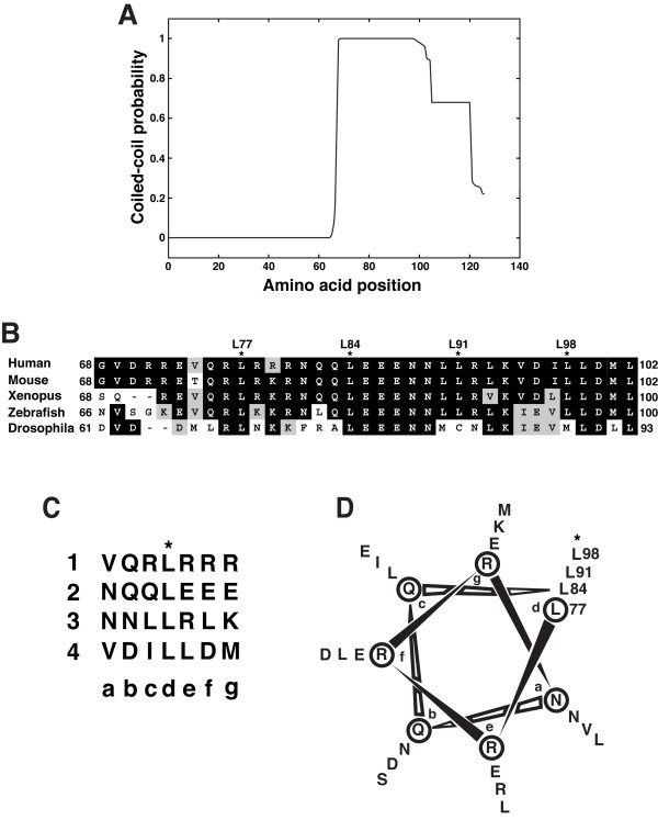
Background: The Wnt/beta-catenin signaling pathway plays crucial roles in embryonic development and in maintenance of organs and tissues in adults. Chibby (Cby) is an evolutionarily conserved molecule that physically interacts with the key downstream coactivator beta-catenin and represses its transcriptional activation potential. Although Cby harbors a predicted coiled-coil motif in the C-terminal region, its molecular nature and functional importance remain largely unexplored.

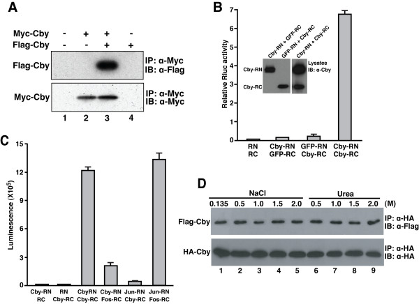
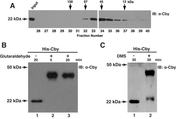
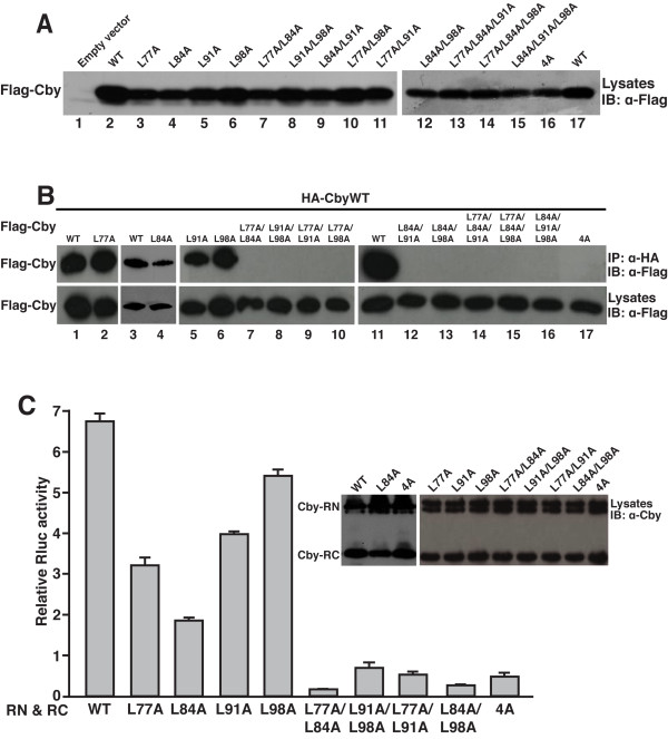
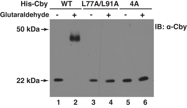
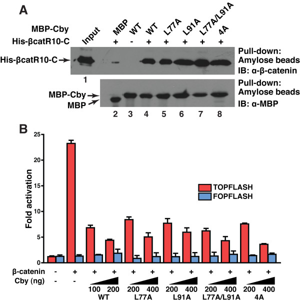
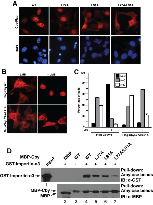
Results: Here we report that Cby forms a stable complex with itself. Alanine substitutions of two or more of four critical leucine residues within the C-terminal heptad repeats completely eliminate the Cby-Cby interaction. The Cby oligomer predominantly exists as a homodimer. Furthermore, we found that dimerization-deficient Cby mutants still retain the ability to bind to beta-catenin and to repress beta-catenin-dependent gene activation. More importantly, Cby homodimerization is required for its efficient interaction with the nuclear import receptor importin-alpha and subsequent nuclear translocation.

Conclusion: Our comprehensive mutational analysis of the Cby coiled-coil domain reveals that the four heptad leucine residues play an essential role in mediating Cby homodimerization. Although monomeric Cby is sufficient to bind to beta-catenin and block beta-catenin-mediated transcriptional activation, homodimer formation of Cby is indispensable for its efficient nuclear import.


Figures

Figure 1
Figure 1
Cby forms a stable complex with itself. (A) Coimmunoprecipitation of Myc-Cby and Flag-Cby. (B) Cby self-interaction was detected by split synthetic Renilla luciferase (hRluc) protein-fragment-assisted complementation. Cby or negative control GFP was fused in-frame to the N-terminal portion (RN) and C-terminal portion (RC) of hRluc. These expression plasmids (400 ng each) were transfected into HEK293T cells as indicated, and Renilla luciferase (Rluc) activities were measured 24 hr post-transfection. A firefly luciferase plasmid (5 ng) was co-transfected to normalize transfection efficiency. Transfections were carried out in triplicate and the means ± SD are shown.(C) Cby-Cby interactions were detectable in vivo in real time using cell-permeable ViviRen live cell substrate. Cby-RN, Cby-RC, Fos-RC and Jun-RN were transiently expressed in HEK293T cells as shown, and ViviRen was added to the tissue culture media 24 hr post-transfection for luminescence measurements. Transfections were carried out in triplicate and the means ± SD are shown.(D) Cby complex is highly stable. The immunoprecipitates were washed three times with wash buffer containing 0.135, 0.5, 1.0, 1.5 or 2.0 M NaCl, or 0.5, 1.0, 1.5 or 2.0 M urea as shown. The experiments with urea were performed in the presence of 0.135 M NaCl.
Figure 2
Figure 2
Cby exists as a homodimer. (A) Gel filtration analysis of Cby. His-Cby purified from stable HEK293T cells was run on a pre-calibrated Superdex 75 FPLC column in buffer containing 1 M NaCl. An aliquot of each fraction was resolved by SDS-PAGE and analyzed by Western blotting using anti-Cby antibody. The arrows indicate the elution positions of protein standards: aldolase, 158 kDa; bovine serum albumin, 67 kDa; ovalbumin, 45 kDa and cytochrome C, 12 kDa. Similar results were obtained using buffer containing 0.5 M NaCl (data not shown). (B, C) Cross-linking experiments. His-tagged Cby was purified from transiently transfected HEK293T cells using Ni-NTA beads, and subjected to cross-linking with glutaraldehyde for 5 or 20 min (B), or dimethyl suberimidate (DMS) for 30 min (C). The samples were then resolved by SDS-PAGE, followed by immunoblotting with anti-Cby antibody.
Figure 3
Figure 3
The C-terminal region of Cby harbors a putative coiled-coil motif. (A) The human Cby protein sequence (126 amino acid residues) was analyzed by the COILS program at a window size of 21 residues. Note that high coiled-coil probabilities exceeding 0.9 were evident from amino acid 68 to 102. (B) Sequence alignment of the Cby coiled-coil domain across species. Identical and similar residues are highlighted in black and gray, respectively. Four leucines, indicated by asterisks, appear at every seventh position, indicative of a leucine-zipper motif. (C) The putative coiled-coil motif of human Cby is depicted as heptad repeats of seven amino acids. The letters a through g designate the positions of residues within the heptad with hydrophobic residues generally found at the a and d positions. The four consecutive heptads are numbered 1 through 4. Note that the d positions are occupied by leucine residues.(D) Helical wheel diagram of the coiled-coil domain of human Cby (amino acids 77–98).
Figure 4
Figure 4
The heptad leucine residues within the coiled-coil domain are crucial for Cby homodimerization. (A) Expression levels of Cby point mutants. Lysates from HEK293T cells transfected with an equal amount (600 ng) of an expression plasmid for Flag-tagged wild-type (WT) or mutant Cby were subjected to Western blotting with an anti-Flag antibody. (B) Cell lysates were prepared from HEK293T cells transiently co-transfected with HA-CbyWT and Flag-CbyWT or individual Cby variants with all possible combinations of leucine-to-alanine mutations [single, double, triple and quadruple (4A)], and subjected to immunoprecipitation with anti-HA antibody. The immunoprecipitates were separated by SDS-PAGE and immunoblotted with anti-Flag antibody. To ensure sufficient protein expression levels, the amounts of plasmid DNA expressing Flag-tagged Cby mutants were increased by 2-fold for transfection, compared to those of Flag-CbyWT plasmid. (C) Split hRluc protein-fragment-assisted complementation assays using Cby point mutants. CbyWT or each of the indicated Cby mutants was fused in-frame to RN and RC. These expression plasmids (400 ng each) were transfected into HEK293T cells, and Rluc activities were measured as described in the legend to Figure 1B. Transfections were carried out in triplicate and the means ± SD are shown. Immunoblotting with anti-Cby antibody showed that the fusion proteins were stably expressed. Cby-RN fusion proteins appear as a doublet probably due to degradation.
Figure 5
Figure 5
Glutaraldehyde cross-linking of dimerization-deficient Cby mutants. His-tagged CbyWT, L77A/L91A or 4A was transiently expressed in HEK293T cells, and purified using Ni-NTA beads. The purified Cby protein was incubated in the absence or presence of glutaraldehyde for 5 min, and resolved on SDS-PAGE, followed by immunoblotting with anti-Cby antibody.
Figure 6
Figure 6
Cby homodimerization is dispensable for its interaction with β-catenin and for repression of β-catenin signaling activity. (A) Binding of Cby point mutants to β-catenin was evaluated by in vitro pull-down assays. Bacterially produced MBP or individual MBP-Cby protein was incubated with His-tagged β-catenin C-terminal domain (His-βcatR10-C). The protein complexes were then pulled down with amylose resin and subjected to Western blotting using anti-β-catenin antibody (top panel). The input lane was loaded with one-fiftieth of the amount of His-βcatR10-C used in the binding reactions (lane 1). One-thirtieth of each pull-down sample was run on a separate SDS-PAGE and immunoblotted with anti-MBP antibody, showing that similar amounts of MBP-Cby protein were pulled down (bottom panel). (B) The ability of Cby mutants to repress β-catenin signaling was tested by TOPFLASH assays. HEK293T cells were transfected with 60 ng of TOPFLASH or mutant FOPFLASH luciferase reporter, with or without 40 ng of an expression vector for stabilized β-catenin (β-catenin-Myc), and the indicated amounts of a Flag-tagged Cby expression vector. Luciferase activity was measured 24 hr post-transfection, and normalized to Renila luciferase activity used as an internal control. Transfections were carried out in triplicate and the means ± SD are shown. Note that, to compensate protein levels, higher amounts of plasmid DNA for the Cby mutants were used for transfection.
Figure 7
Figure 7
Homodimer formation of Cby is a prerequisite for its efficient nuclear import. (A) C-terminally Flag-tagged CbyL77A/L91A accumulates in the cytoplasm. COS7 cells were transiently transfected with an expression vector encoding C-terminally Flag-tagged WT or mutant Cby as indicated. Cells were fixed 24 hr post-transfection, followed by immunostaining with anti-Flag antibody. Nuclei were stained with DAPI. (B) A fraction of N-terminally Flag-tagged CbyL77A/L91A is found in the cytoplasm after LMB treatment. COS7 cells were transfected with an expression vector for N-terminally Flag-tagged CbyWT or CbyL77A/L91A, treated with methanol (- LMB) or 40 nM LMB (+ LMB) for 5 hr, and immunostained with anti-Flag antibody for Cby. Nuclei were counterstained with DAPI. (C) Quantitative analysis of the results in (B). The subcellular localization of Flag-CbyWT and Flag-CbyL77A/L91A was scored as follows: N>C, predominantly nuclear; N = C, evenly distributed between the nucleus and cytoplasm; N<C, predominantly cytoplasmic. Error bars represent the means ± SD of three independent experiments. (D) Reduced binding of CbyL77A/L91A to importin-α. Bacterially produced MBP or the indicated MBP-Cby protein was incubated with GST-importin-α3. The complexes were then pulled down with amylose resin and subjected to Western blotting using anti-GST antibody (top panel). The input lane was loaded with one-fiftieth of the amount of GST-importin-α3 used in the binding reactions (lane 1). One-thirtieth of each pull-down sample was run on a separate SDS-PAGE and immunoblotted with anti-MBP antibody, showing that similar amounts of MBP-Cby protein were pulled down (bottom panel).

Similar articles

Cited by

References

    1. Wodarz A, Nusse R. Mechanisms of Wnt signaling in development. Annu Rev Cell Dev Biol. 1998;14:59–88. - PubMed
    1. Miller JR. The Wnts. Genome Biol. 2002;3:REVIEWS3001. - PMC - PubMed
    1. Pinto D, Clevers H. Wnt control of stem cells and differentiation in the intestinal epithelium. Exp Cell Res. 2005;306:357–363. - PubMed
    1. Nusse R. Wnt signaling in disease and in development. Cell Res. 2005;15:28–32. - PubMed
    1. Moon RT, Kohn AD, De Ferrari GV, Kaykas A. WNT and beta-catenin signalling: diseases and therapies. Nat Rev Genet. 2004;5:691–701. - PubMed

Publication types

MeSH terms

LinkOut - more resources