. 2009 Jun 1;122(Pt 11):1852-62.
doi: 10.1242/jcs.046870. Epub 2009 May 12.

A FAK-p120RasGAP-p190RhoGAP complex regulates polarity in migrating cells

Affiliations

A FAK-p120RasGAP-p190RhoGAP complex regulates polarity in migrating cells

Alok Tomar et al. J Cell Sci. .

Erratum in

  • J Cell Sci. 2009 Aug 15;122(Pt 16):3005

Abstract

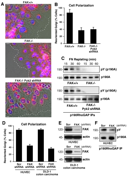
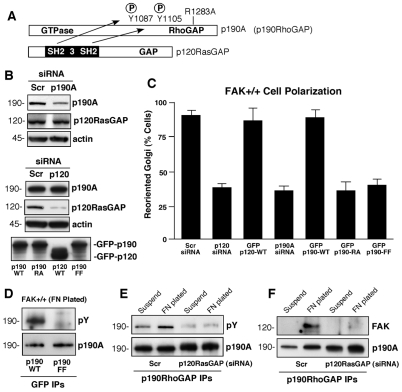
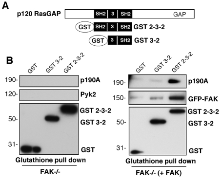
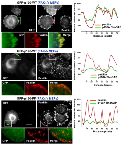
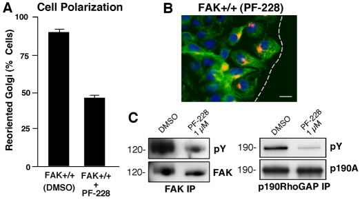
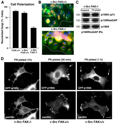
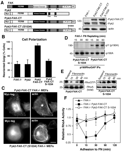
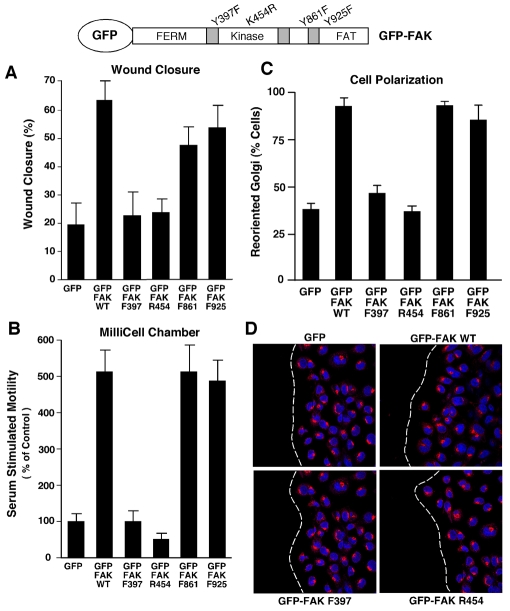
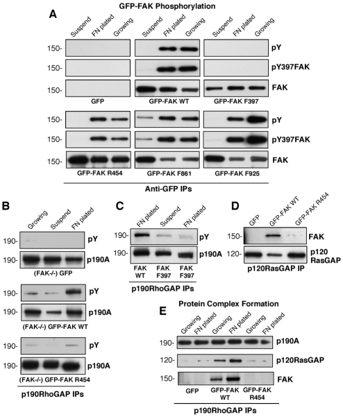
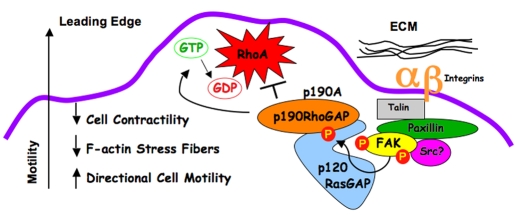
Directional motility is a complex process requiring the spatiotemporal integration of signals that regulate cytoskeletal changes, and the establishment of an anteroposterior or polarized cell axis. Focal adhesion kinase (FAK) promotes cell migration, but a molecular role for FAK in promoting cell polarity remains undefined. Here, using wound healing and Golgi-reorientation analyses, we show that fibroblast, endothelial and carcinoma polarity during cell migration requires FAK and is associated with a complex between FAK, p120RasGAP and p190RhoGAP (p190A), leading to p190A tyrosine phosphorylation. Fibronectin-integrin-mediated FAK activation and phosphorylation promote SH2-mediated binding of p120RasGAP to FAK and FAK-mediated p190A tyrosine phosphorylation. The association of p120RasGAP with FAK facilitates the formation of a FAK-p120RasGAP-p190A complex targeted to leading-edge focal adhesions by FAK. Knockdown of p120RasGAP, mutation of FAK Y397 or inhibition of FAK activity prevent the association of FAK with p190A and subsequent tyrosine phosphorylation of p190A, and result in the loss of cell polarity. Because reconstitution of FAK-null fibroblasts with FAK or a Pyk2-FAK chimera restore the normal decrease in RhoA GTP binding upon cell spreading on fibronectin, our studies support a model whereby FAK activity facilitates the recruitment and stabilization of a p120RasGAP-p190A complex at leading-edge focal adhesions connected to the transient inhibition of RhoA activity and the regulation of cell polarity.


Figures

Fig. 1.
Fig. 1.
FAK promotes cell polarity and is required for p190A tyrosine phosphorylation after FN plating. (A) FAK+/+, FAK–/– and FAK–/– Pyk2-shRNA MEFs were grown on FN-coated coverslips. Cells were wounded and allowed to migrate in the presence of serum for 4 hours. Cells were fixed, imaged in phase, and stained for Golgi (β-Cop, red) and nuclear (Hoechst, blue) markers. The position of the leading lamella (broken line) is indicated. Scale bars: 15 μm. (B) Golgi reorientation analyses, a square was drawn over cell nuclei at the wound edge and divided into quadrants. Reoriented Golgi was scored positive by β-Cop staining entirely within the quadrant facing the leading edge. In total, 100 cells were analyzed; data is percent of cells analyzed ± s.d. (C) p190A immunoprecipitations (IPs) were made from FAK–/–, FAK+/+ and FAK–/– Pyk2-shRNA lysates of serum-starved cells held in suspension for 1 hour and replated onto FN-coated dishes for the indicated times. p190A IPs were sequentially analyzed by anti-phosphotyrosine (pY) and anti-p190A immunoblotting. (D) HUVEC and DLD-1 carcinoma cells expressing scrambled (Scr) or anti-FAK shRNA were analyzed for Golgi reorientation after scratch wounding as above. In total, 100 cells lining the wound edge were scored (± s.d.). (E) FAK expression is reduced by anti-FAK compared with Scr shRNA as determined by immunoblotting. (F) p190A IPs from lysates from HUVECs expressing Scr or anti-FAK shRNA that were replated onto FN-coated dishes (30 minutes) and analyzed by anti-phosphotyrosine (pY) and anti-p190A immunoblotting.
Fig. 2.
Fig. 2.
p120RasGAP promotes FN-stimulated p190A tyrosine phosphorylation, FAK association with p190A and cell polarity. (A) Schematic of the p190A and p120RasGAP proteins, and p120RasGAP SH2-mediated binding to phosphorylated Y1087 and Y1105 in p190A. R1283A (RA) mutation disrupts p190A GAP activity. (B) Scrambled (Scr)-, p190A- or p120RasGAP-siRNA transfection of MEFs and the associated protein levels after 48 hours. Actin levels were used as a control. Anti-GFP immunoblotting was used to confirm transient GFP–p190-WT, GFP–p190-RA, GFP–190-FF (Y1087F, Y1105F) and GFP-p120RasGAP expression. (C) Golgi reorientation after scratch wounding was performed with Scr p190A siRNA or p120RasGAP siRNA with siGlo co-transfection as a marker to detect transfected MEFs. Data is the percentage of 100 cells analyzed and is combined with similar analyses of MEFs transfected with GFP-p120RasGAP, GFP-p190A-WT, GFP-p190A-RA, or GFP-p190A-FF. Values are from 100 cells ± s.d. (D) Mutation of p190A Y1087F and Y1105F (p190A-FF) blocks FN-stimulated tyrosine phosphorylation as determined by transfection, anti-GFP-tag immunoprecipitation (IP), and sequential anti-pY and anti-HA immunoblotting. (E) p120RasGAP siRNA-knockdown disrupts p190A tyrosine phosphorylation. Lysates from MEFs in suspension or FN plated (30 minutes) were analyzed by anti-p190A IPs followed by anti-pY and anti-p190A immunoblotting. (F) p120RasGAP siRNA-knockdown disrupts FAK-p190A association upon FN plating. Lysates from MEFs in suspension or FN plated (30 minutes) were analyzed by anti-p190A IPs followed by anti-FAK and anti-p190A immunoblotting.
Fig. 3.
Fig. 3.
The p120RasGAP SH2-SH3-SH2 (2-3-2) region bridges FAK and p190A. (A) Schematic representation of the mammalian GST expression vectors of GST–2-3-2 and GST–3-2 of p120RasGAP. (B) Coexpression of GST–2-3-2 and FAK facilitate the formation of a complex with p190A. FAK–/– MEFs were transiently transfected with the indicated GST constructs (left) or GST constructs plus GFP-FAK (right), plated (30 minutes) onto FN-coated dishes and lysates were analyzed by glutathione-agarose bead pull down. Association of endogenous Pyk2, p190A or GFP-FAK was determined by immunoblotting.
Fig. 4.
Fig. 4.
FAK and p190A tyrosine phosphorylation promote p190A FA localization upon FN plating. FAK+/+ or FAK–/– MEFs were transfected with GFP–p190-WT or GFP–p190-FF, serum starved, plated onto FN (10 μg/ml, 30 minutes), and analyzed for GFP and paxillin colocalization. Fluorescence-intensity profiles represent the area marked by the colored lines (green p190A, red paxillin). Insets, enlargement of the area of interest shown for GFP-p190A, paxillin and the merged images. GFP–p190-WT and paxillin show colocalization at peripheral FAs in FAK+/+ but not in FAK–/– MEFs as determined by fluorescent-intensity overlap. The GFP–p190-FF fluorescent-intensity profile does not overlap with paxillin at FAs. Scale bar: 20 μm.
Fig. 5.
Fig. 5.
Pharmacological FAK inhibition blocks MEF polarization and p190A tyrosine phosphorylation. (A) Golgi-reorientation analyses of scratch-wounded FAK+/+ MEFs performed in the presence of DMSO or 1 μM PF-228 (FAK inhibitor). Data is the percentage of 100 cells with Golgi that were reoriented towards the wound edge (± s.d.). (B) Representative image from a wound healing assay. MEFs were stained with β-Cop antibody (red), anti-tubulin antibody (green) and Hoechst nuclear dye (blue). The position of leading lamella (broken white line) is shown. Scale bar: 30 μm. (C) FAK and p190A IPs from FN-plated MEFs (30 minutes) in the presence of DMSO or PF-228 (1 μM) were sequentially analyzed by anti-pY followed by anti-FAK or anti-p190A immunoblotting, respectively.
Fig. 6.
Fig. 6.
FAK is required for Src-transformed MEF polarity independent of p190A tyrosine phosphorylation. (A) Scratch-wound-healing Golgi-reorientation analyses performed with normal FAK+/+ or Src-transformed FAK+/+ and FAK–/– MEFs. Data is the percentage of 100 cells with Golgi that were reoriented towards the wound edge (± s.d.). Representative images from a wound healing assay. Src FAK+/+ and Src FAK–/– MEFs were stained with β-Cop antibody (red), anti-tubulin antibody (green) and Hoechst nuclear dye (blue). The position of leading lamella (broken white line) is shown. Scale bar: 30 μm. (C) Src promotes adhesion-independent and FAK-independent p190A tyrosine phosphorylation. p190A IPs from suspended or FN-replated Src FAK–/– MEF lysates analyzed for p190A tyrosine phosphorylation and p120RasGAP association by anti-pY, anti-p120RasGAP and anti-p190A immunoblotting. (D) FAK facilitates p190A membrane and FA localization upon Src MEF adhesion to FN. Src FAK–/– or Src FAK+/+ MEFs were transfected with GFP–p190A-WT and plated onto FN-coated coverslips for 30 minutes or 1 hour as indicated. Cells were imaged for GFP-p190A and co-stained for paxillin localization (rhodamine). Arrows show the localization of p190A at membrane projections at 30 minutes and at peripheral FAs at 1 hour in Src FAK+/+ MEFs. GFP-p190A exhibits a punctate distribution in Src FAK–/– MEFs. Scale bar: 10 μm.
Fig. 7.
Fig. 7.
Pyk2 that is targeted to FAs can substitute for FAK in promoting MEF polarity, p190A tyrosine phosphorylation and RhoA regulation. (A) Schematic representation of HA-FAK (murine), Myc-Pyk2 (human), Pyk2–FAK-CT (Pyk2 residues 1-682 including the kinase domain fused to FAK residues 680-1052) and Pyk2–FAK-CT-S1034 [containing a point mutation within the FAK FA-targeting (FAT) domain that disrupts paxillin binding]. Expression levels of endogenous Pyk2 and Pyk2–FAK-CT constructs within FAK–/– MEFs determined by anti-Pyk2 and anti-actin immunoblotting are shown on the right. (B) Scratch-wound-healing Golgi-reorientation analyses in FAK–/– MEFs and FAK–/– MEFs expressing Pyk2, Pyk2–FAK-CT or Pyk2–FAK-CT-S1034. Data is the percentage of 100 cells with Golgi that are reoriented towards the wound edge (± s.d.). (C) Disruption of Pyk2–FAK-CT FA localization that is associated with S1034 mutation. Indirect immunofluorescent localization of Pyk2–FAK-CT and Pyk2–FAK-CT-S1034 by anti-Myc-tag (fluorescein) and actin (phalloidin) staining of FN-plated (45 minutes) MEFs is shown. Scale bar: 10 μm. (D) The S1034 mutation disrupts Pyk2–FAK-CT-facilitated p190A tyrosine phosphorylation. p190A IPs, made from Pyk2–FAK-CT and Pyk2–FAK-CT-S1034 FAK–/– MEFs that were replated onto FN-coated dishes for the indicated times, were sequentially analyzed by anti-pY and anti-p190A blotting. (E) Pyk2–FAK-CT expression results in less Rho GTPase activation upon FN plating. GTP-bound RhoA levels were determined by affinity pulldown assays from lysates of suspended and FN-replated FAK–/– Pyk2–FAK-CT and FAK–/– Pyk2–FAK-CT-S1034 MEFs followed by blotting for total RhoA levels. (F) Quantitation of FN-associated RhoA-GTP binding. Data is plotted as relative RhoA-GTP levels in suspended FAK–/– MEFs and is the mean of two independent experiments. Error bars show ± s.d.
Fig. 8.
Fig. 8.
FAK Y397 phosphorylation and kinase activity are required for directional MEF motility and polarization. Schematic representation of mutations within GFP-FAK constructs used to reconstitute FAK–/– MEFs (top panel). (A) WT- and mutant-FAK-reconstituted MEFs were grown to confluence, serum starved for 16 hours, scratch-wounded using a pipette tip and allowed to migrate for 20 hours in the presence of serum. The percentage of wound closure was calculated from three independent experiments, presented ± s.d. (B) Directional motility of WT- and mutant-FAK-reconstituted MEFs was measured using FN-coated (10 μg/ml) Boyden-chamber (top and bottom) motility over 4 hours. Serum-containing media was added to the lower chamber. The number of motile cells (membrane underside) from three independent experiments is expressed as the percentage of FAK–/– GFP control (normalized to 100, ± s.d.). (C) Golgi-reorientation analyses of GFP-expressing and GFP-FAK-reconstituted FAK–/– MEFs. Data is the percentage of cells that had reoriented Golgi towards the wound edge (100 cells ± s.d.). (D) Representative images of scratch-wounded GFP-expressing or indicated GFP-FAK-reconstituted MEFs after 4 hours as analyzed by β-Cop Golgi (rhodamine; red) and Hoechst nuclear (blue) staining. The position of the leading lamella (broken white line) is shown.
Fig. 9.
Fig. 9.
FAK activity facilitates p190A tyrosine phosphorylation and complex formation with p120RasGAP and p190A. (A) Regulation of GFP-FAK phosphorylation in reconstituted FAK–/– MEFs. Lysates were made from starved cells held in suspension (1 hour), cells that were replated onto FN-coated dishes (45 minutes) and from growing cells. Anti-GFP IPs were sequentially analyzed by anti-pY, FAK pY397 phospho-specific and total FAK blotting. (B) p190A tyrosine phosphorylation is dependent upon FAK activity. p190A IPs were made from growing, suspended or FN-plated (30 minutes) FAK–/– MEFs expressing GFP or reconstituted with GFP–FAK-WT or GFP–FAK-R454 and analyzed by anti-pY followed by anti-p190A immunoblotting. (C) Mutation of FAK Y397 blocks p190A tyrosine phosphorylation. p190A IPs from suspended or FN-plated FAK–/– MEFs expressing GFP–FAK-WT or GFP–FAK-F397 were analyzed by anti-pY followed by anti-p190A immunoblotting. (D) FAK activity is required for p120RasGAP association. p120RasGAP IPs from FN-plated FAK–/– MEFs expressing GFP or reconstituted with GFP–FAK-WT or GFP–FAK-R454 were analyzed for FAK association by anti-FAK followed by anti-p120RasGAP immunoblotting. (E) p190A-p120RasGAP-FAK association is dependent upon FN-stimulated FAK activity. p190A IPs from growing or FN-plated FAK–/– MEFs expressing GFP or reconstituted with GFP–FAK-WT or GFP–FAK-R454 were analyzed for p120RasGAP and FAK association by sequential immunoblotting.
Fig. 10.
Fig. 10.
Model of the spatial and temporal regulation of p190A by FAK. FAK is activated by integrin clustering and is localized to leading-edge FAs in migrating MEFs via binding to the integrin-associated proteins paxillin and talin. FAK Y397 phosphorylation promotes the SH2-mediated binding of p120RasGAP to FAK and the SH2-SH3-SH2 region of p120RasGAP facilitates a bridge between FAK and p190A. Increased p190A tyrosine phosphorylation is dependent upon FN-stimulated FAK activity that can either phosphorylate p190A directly (Holinstat et al., 2006) or promote p190A tyrosine phosphorylation via Src activation (Wu et al., 2008). In cells with activated Src, cell polarity and recruitment of p190A to leading-edge paxillin-containing FAs is dependent upon FAK expression. Increased GAP activity of tyrosine-phosphorylated p190A that is localized to FAs acts to inhibit RhoA GTPase activity, and this is associated with decreased actin contractility at leading-edge lamella and enables directional or polarized cell movement.

References

    1. Arthur, W. T. and Burridge, K. (2001). RhoA inactivation by p190RhoGAP regulates cell spreading and migration by promoting membrane protrusion and polarity. Mol. Biol. Cell 12, 2711-2720. - PMC - PubMed
    1. Arthur, W. T., Petch, L. A. and Burridge, K. (2000). Integrin engagement suppresses RhoA activity via a c-Src-dependent mechanism. Curr. Biol. 10, 719-722. - PubMed
    1. Bass, M. D., Morgan, M. R., Roach, K. A., Settleman, J., Goryachev, A. B. and Humphries, M. J. (2008). p190RhoGAP is the convergence point of adhesion signals from alpha 5 beta 1 integrin and syndecan-4. J. Cell Biol. 181, 1013-1026. - PMC - PubMed
    1. Beardsley, A., Fang, K., Mertz, H., Castranova, V., Friend, S. and Liu, J. (2005). Loss of caveolin-1 polarity impedes endothelial cell polarization and directional movement. J. Biol. Chem. 280, 3541-3547. - PubMed
    1. Bernards, A. and Settleman, J. (2004). GAP control: regulating the regulators of small GTPases. Trends Cell Biol. 14, 377-385. - PubMed

Publication types

MeSH terms