. 2009 Jun 1;122(Pt 11):1882-94.
doi: 10.1242/jcs.039057. Epub 2009 May 12.

VASP is a CXCR2-interacting protein that regulates CXCR2-mediated polarization and chemotaxis

Affiliations

VASP is a CXCR2-interacting protein that regulates CXCR2-mediated polarization and chemotaxis

Nicole F Neel et al. J Cell Sci. .

Abstract

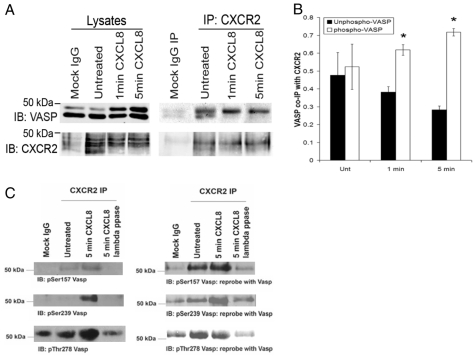
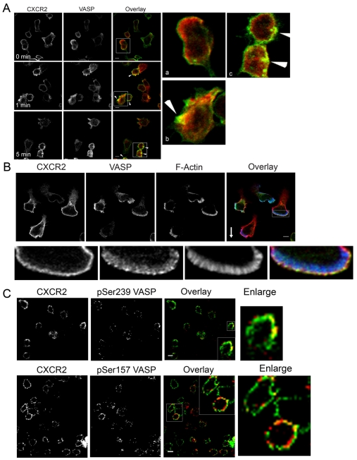
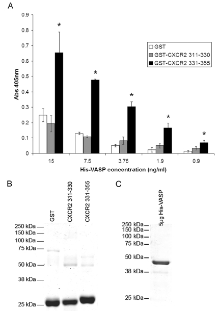
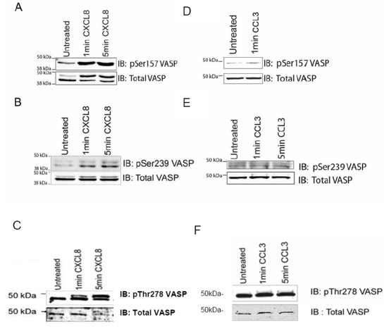
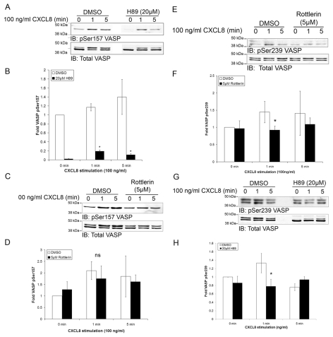
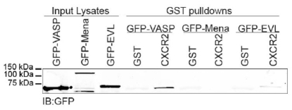
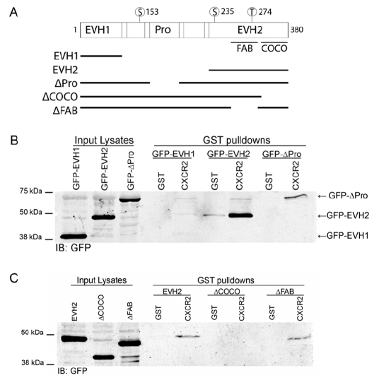
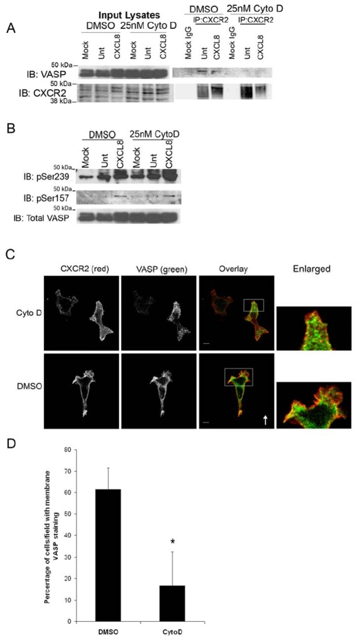
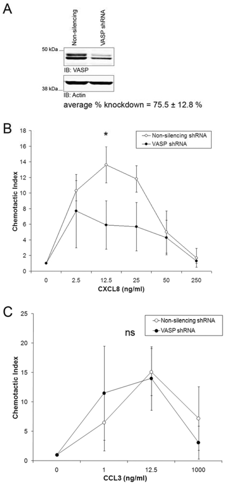
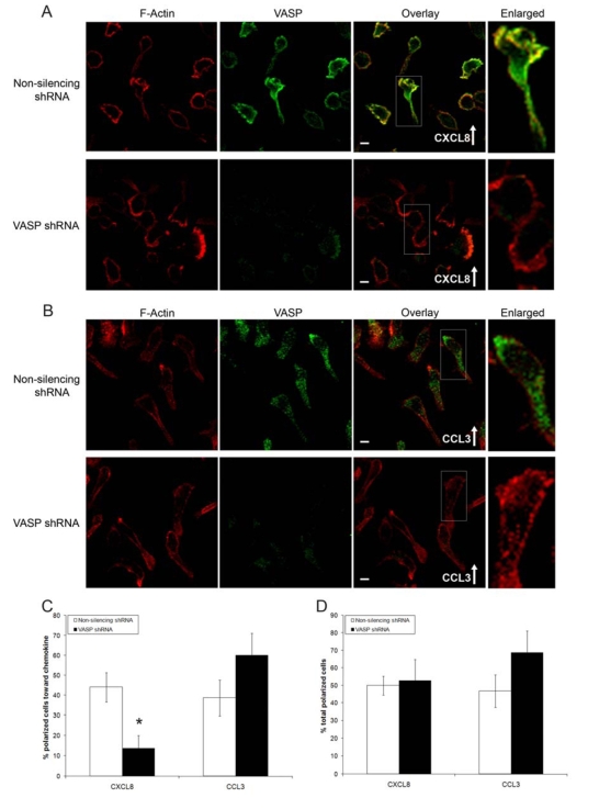
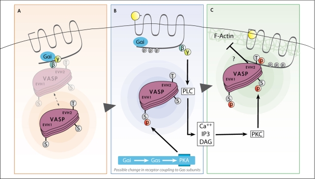
Chemotaxis regulates the recruitment of leukocytes, which is integral for a number of biological processes and is mediated through the interaction of chemokines with seven transmembrane G-protein-coupled receptors. Several studies have indicated that chemotactic signaling pathways might be activated via G-protein-independent mechanisms, perhaps through novel receptor-interacting proteins. CXCR2 is a major chemokine receptor expressed on neutrophils. We used a proteomics approach to identify unique ligand-dependent CXCR2-interacting proteins in differentiated neutrophil-like HL-60 cells. Using this approach, vasodilator-stimulated phosphoprotein (VASP) was identified as a CXCR2-interacting protein. The interaction between CXCR2 and VASP is direct and enhanced by CXCL8 stimulation, which triggers VASP phosphorylation via PKA- and PKCdelta-mediated pathways. The interaction between CXCR2 and VASP requires free F-actin barbed ends to recruit VASP to the leading edge. Finally, knockdown of VASP in HL-60 cells results in severely impaired CXCR2-mediated chemotaxis and polarization. These data provide the first demonstration that direct interaction of VASP with CXCR2 is essential for proper CXCR2 function and demonstrate a crucial role for VASP in mediating chemotaxis in leukocytes.


Figures

Fig. 1.
Fig. 1.
CXCR2 and VASP interact in dHL-60-CXCR2 cells. (A) Lysates from cells stimulated with vehicle (Mock IgG, Untreated) or cells stimulated with 100 ng/ml CXCL8 for 1 or 5 minutes were incubated with normal rabbit IgG-(Mock IgG) or rabbit anti-CXCR2 antibody-coupled Sepharose. Immunoprecipitated proteins were eluted and analyzed by SDS-PAGE and western blot (IB) for CXCR2 and VASP. (B) Quantification of unphosphorylated and Ser157-phosphorylated VASP coimmunoprecipitating with CXCR2. Values are normalized to total VASP levels and represented as mean ± s.e.m. Significant differences between the unphosphorylated and phosphorylated VASP in the IP are indicated (*P≤0.05, Mann Whitney U-test). (C) dHL-60-CXCR2 cells not treated (lane 2) or treated (lane 3) with 100 ng/ml CXCL8 for 5 minutes were lysed, subjected to immunoprecipitation as above, and analyzed by SDS-PAGE and western blot (IB) for Ser157-P, Ser239-P or P-Thr278-P VASP using ECL, or for total VASP using the Odyssey system or ECL. One set of immunoprecipitated lysates stimulated with CXCL8 was incubated with lambda phosphatase (lane 4) as described in Materials and Methods. Data shown are representative of three separate experiments.
Fig. 2.
Fig. 2.
CXCR2 and VASP localize to plasma membrane ruffles upon global and directional CXCL8 stimulation. (A) Immunofluorescence confocal images of CXCR2 and VASP staining in dHL-60-CXCR2 cells stimulated globally with vehicle (0 minutes) or 100 ng/ml of CXCL8 for 1 or 5 minutes. Colocalization after both 1 and 5 minutes of stimulation is indicated by arrows. Images represent single z-sections of 0.38 μm. Overlay images are pseudocolored where green is CXCR2 and red is VASP. Images after 0 minutes (a), 1 minute (b), and 5 minutes (c) stimulation are enlarged ×5 from the boxed areas on the left. (B) Immunofluorescence confocal images of CXCR2, VASP and F-actin staining in dHL-60-CXCR2 cells stimulated directionally with 50 ng/ml CXCL8 in a Zigmond chamber for 15 minutes. The arrow indicates the direction of CXCL8 gradient (0-50 ng/ml CXCL8). Image represents a single z-section of 0.35 μm pseudocolored, where green is VASP, red is CXCR2, and blue is F-actin. (C) Immunofluorescence confocal images of CXCR2 and VASP phosphorylated on Ser157 (pPSer157 VASP) or Ser239 (pSer239) following 1 minute of global CXCL8 (100 ng/ml) stimulation. Overlay images are pseudocolored, where green is CXCR2 and red is pVASP. Image represents a single z-section of 0.28 μm. Insets are enlarged ×2 from boxed areas in originals. Images were processed using AdobePhotoshop. Scale bars: 5 μm. Data shown are representative of three separate experiments.
Fig. 3.
Fig. 3.
Purified His6-VASP interacts with amino acids 331-355 from the CXCR2 C-terminus. (A) 96-well plates were coated with 3 μg/ml GST, GST-CXCR2 311-330, or GST-CXCR2 331-355 and incubated with various concentrations of His6-VASP (0.06-60 ng/ml). Bound His6-VASP was detected using nickel-activated HRP (Abs 405 nm). Triplicate experiments show His6-VASP binding (mean ± s.e.m.) to GST-CXCR2 331-355 was statistically higher than to GST alone or GST-CXCR2 311-330 (*P≤0.05, Mann Whitney U-test). Data shown are representative of at least three separate experiments. (B) Representative Coomassie-blue-stained gel of 5 μg purified GST, GST-CXCR2 311-330 and GST-CXCR2 331-355. (C) Representative Coomassie-blue-stained gel of 5 μg purified His6-VASP.
Fig. 4.
Fig. 4.
VASP is phosphorylated on Ser157, Ser239 and Thr278 specifically in response to CXCL8 stimulation. HL-60-CXCR2 cells were stimulated with vehicle (Untreated) or 100 ng/ml CXCL8 for 1 or 5 minutes. Lysates were subjected to SDS-PAGE and western blot analysis (IB) using antibodies specific for Ser157-P (A), Ser239-P (B) or Thr278-P VASP (C). HL-60-CXCR2 cells were stimulated with vehicle (Untreated) or 100 ng/ml CCL3 for 1 minute. Lysates were subjected to SDS-PAGE and western blot analysis (IB) for Ser157-P (D), Ser239-P (E) or Thr278-P VASP (F). Data shown are representative of three separate experiments.
Fig. 5.
Fig. 5.
Phosphorylation of VASP upon CXCL8 stimulation is mediated through PKA and PKC. Lysates from HL-60-CXCR2 cells pretreated with various inhibitors, stimulated with vehicle (0 minutes) or 100 ng /ml CXCL8 for 1 or 5 minutes and analyzed by SDS-PAGE and western blot. Statistical difference of mean ± s.e.m. in DMSO versus treatment is indicated (*P≤0.05, Mann Whitney U-test). (A) Cells pretreated with DMSO or 20 μM H89 for 60 minutes and analyzed by western blot using antibodies specific for VASP and VASP Ser157-P. (B) Quantification of normalized density of VASP Ser157-P in DMSO-versus H89-treated samples. (C) Cells pretreated with DMSO or 5 μM rottlerin for 15 minutes and analyzed by western blot analysis for VASP and VASP Ser157-P. (D) Quantification of normalized density of VASP Ser157-P in DMSO-versus rottlerin-treated samples. (E) Cells pretreated with DMSO or 5 μM rottlerin for 15 minutes and analyzed for VASP and VASP Ser239-P (F). Quantification of normalized density of VASP Ser239-P in DMSO-versus rottlerin-treated samples. (G) Cells pretreated with DMSO or 20 μM H89 for 60 minutes and analyzed using antibodies for VASP and VASP Ser239-P. (H) Quantification of normalized density of VASP Ser239-P in DMSO-versus H89-treated samples. Data shown represent average quantification from at least three separate experiments.
Fig. 6.
Fig. 6.
Phosphorylation of VASP regulates its interaction with CXCR2. (A) Western blot analysis for GFP (IB) of GST pull-downs with GST or GST-CXCR2 C-terminus using lysates from MVD7 cells stably expressing GFP-VASP WT, S153A-S235A, S153-T274A and S235A-T274A. (B) Western blot analysis of purified His6-VASP EVH2 domain incubated with catalytic subunit of PKA with and without ATP using a Ser235-P-specific antibody. (C) Binding (mean ± s.e.m.) of His6-VASP EVH2 domain incubated with and without ATP to either GST or GST-CXCR2 331-355. Binding was detected by measuring absorbance at 405 nm from three separate experiments. Statistical significance of binding to GST-CXCR2 with or without ATP is indicated by *P≤0.05 (Mann Whitney U-test). (D) Representative Coomassie-blue-stained gel of 5 μg purified His6-VASP EVH2. (E) Representative Coomassie-blue-stained gel of 5 μg of purified GST or GST-CXCR2 331-355.
Fig. 7.
Fig. 7.
CXCR2 preferentially interacts with VASP as opposed to Mena or EVL. Western blot analysis for GFP (IB) of GST pull-downs with GST or GST-CXCR2 C-terminus using lysates from MVD7 cells stably expressing GFP-VASP, GFP-Mena or GFP-EVL. Data shown are representative of three separate experiments.
Fig. 8.
Fig. 8.
CXCR2 interaction with VASP occurs through the VASP-EVH2 domain and requires the coiled-coil region. (A) Schematic of murine VASP domain structure and fragments used in pull-down experiments. Numbers indicate the amino acids phosphorylated in murine VASP. Western blot analysis for GFP (IB) to detect GFP-VASP of GST pull-downs with GST or GST-CXCR2 C-terminus using: (B) lysates from MVD7 cells stably expressing GFP-VASP-EVH1, GFP-VASP-EVH2 or GFP-VASP-ΔPro (deletion of proline-rich region) or (C) lysates from MVD7 cells stably expressing GFP-VASP-EVH2 WT, GFP-VASP-ΔCOCO (coiled-coil deletion), or GFP-VASP-ΔFAB (F-actin binding motif deletion). Data shown are representative of three separate experiments.
Fig. 9.
Fig. 9.
F-actin is necessary for localization of VASP to the membrane and interaction with CXCR2. dHL-60 cells were pretreated for 30 minutes with 25 nM cytochalasin D (CytoD) or DMSO (vehicle), then stimulated with vehicle (Mock, Unt) or 100 ng/ml CXCL8 for 1 minute. (A) Cell lysates were incubated with either normal rabbit IgG (Mock IgG) or rabbit anti-CXCR2 antibody-coupled Sepharose. Eluted immunoprecipitated proteins were analyzed by SDS-PAGE and western blot (IB) for CXCR2 and VASP. (B) Western blot analysis (IB) using antibodies specific for VASP P-Ser157 or VASP Ser239-P of lysates. (C) Immunofluorescence confocal images of CXCR2, VASP and F-actin staining in dHL-60-CXCR2 cells and stimulated directionally with 50 ng/ml CXCL8 in Zigmond chamber for 15 minutes. The arrow indicates direction of CXCL8 gradient (0-50 ng/ml CXCL8). Image represents a single z-section of 0.49 μm. Enlarged panel images are magnified ×4 from original images. Overlay image is pseudocolored where green is VASP, red is CXCR2, and blue is F-Actin. Images were processed using Adobe Photoshop. Scale bars: 5 μm. (D) Quantification of mean ± s.e.m. percentage of cells per ×63 field exhibiting VASP immunofluorescence staining at the plasma membrane versus cytoplasm. Ten microscopic fields were examined for DMSO- and CytoD-treated cells with 2-8 cells per field Statistical significance of DMSO-versus CytoD-treated cells is indicated (*P<0.0005, Student's t-test). Data shown are representative of three separate experiments.
Fig. 10.
Fig. 10.
Expression of VASP shRNA specifically impairs CXCR2-mediated chemotaxis. (A) Western blot analysis of VASP in lysates from HL-60-CXCR2 cells stably expressing non-silencing shRNA and VASP shRNA. Average percentage knockdown ± s.e.m. of VASP in western blots from three separate experiments. (B) Boyden chamber assay assessing chemotaxis of HL-60-CXCR2 cells stably expressing non-silencing or VASP shRNA in response to CXCL8 (B) or (C) in response to CCL3. Graphs represent the mean ± s.e.m. chemotactic index from three separate experiments. Statistical difference in dose response curve for CXCL8-mediated chemotaxis in non-silencing versus VASP shRNA expressing cells (*P<0.01, one-way ANOVA). There was no significant difference (ns) in CCL3-mediated chemotaxis between non-silencing versus VASP shRNA expressing cells (one-way ANOVA).
Fig. 11.
Fig. 11.
Cells expressing VASP shRNA exhibit impaired CXCL8-induced cell polarization. Confocal images of cells expressing non-silencing or VASP shRNA stimulated directionally with 50 ng/ml of CXCL8 (A) or 50 ng/ml of CCL3 (B) in a Zigmond chamber for 15 minutes and stained using anti-VASP antibody (green) and phalloidin (red). Arrows indicate the direction of the gradients (0-50 ng/ml CXCL8 or CCL3). Enlarged panels are magnified ×2.5 original images. Scale bars: 5 μm (C). Quantification of mean ± s.e.m. number of cells per field polarized in the direction of the CXCL8 or CCL3 gradients. Statistical significance of non-silencing versus VASP shRNA is indicated (*P<0.001, Mann Whitney U-test). (D) Quantification of the mean ± s.e.m. number of polarized cells from images of 20 ×63 fields, each containing 25-50 cells. Data shown are representative of two separate experiments.
Fig. 12.
Fig. 12.
Model illustrating the roles of VASP phosphorylation in mediating the VASP-CXCR2 interaction. (A) CXCR2 and VASP in unstimulated cells. VASP is predominantly located in the cytoplasm but might have some loose basal association with CXCR2. (B) Upon ligand binding, uncoupling of the G proteins occurs, activating PKA- and PKC-signaling pathways. VASP localizes to the plasma membrane. It is likely that there is a change in Gα coupling to CXCR2 to activate PKA, which phosphorylates VASP on Ser157 in the EVH1 domain. (C) Uncoupling of the Gβγ subunit activates PKC, which phosphorylates Ser239 and possibly Thr278 in the EVH2 domain of VASP. These phosphorylations allow strong interaction between VASP and CXCR2. CXCR2 binding to VASP is likely mutually exclusive with the F-Actin–VASP interaction but it is possible that when phosphorylated VASP is bound to CXCR2, sustained interaction with F-actin occurs.

References

    1. Applewhite, D. A., Barzik, M., Kojima, S., Svitkina, T. M., Gertler, F. B. and Borisy, G. G. (2007). Ena/VASP proteins have an anti-capping independent function in filopodia formation. Mol. Biol. Cell 18, 2579-2591. - PMC - PubMed
    1. Aszodi, A., Pfeifer, A., Ahmad, M., Glauner, M., Zhou, X. H., Ny, L., Andersson, K. E., Kehrel, B., Offermanns, S. and Fassler, R. (1999). The vasodilator-stimulated phosphoprotein (VASP) is involved in cGMP- and cAMP-mediated inhibition of agonist-induced platelet aggregation, but is dispensable for smooth muscle function. EMBO J. 18, 37-48. - PMC - PubMed
    1. Bachmann, C., Fischer, L., Walter, U. and Reinhard, M. (1999). The EVH2 domain of the vasodilator-stimulated phosphoprotein mediates tetramerization, F-actin binding, and actin bundle formation. J. Biol. Chem. 274, 23549-23557. - PubMed
    1. Barzik, M., Kotova, T. I., Higgs, H. N., Hazelwood, L., Hanein, D., Gertler, F. B. and Schafer, D. A. (2005). Ena/VASP proteins enhance actin polymerization in the presence of barbed end capping proteins. J. Biol. Chem. 280, 28653-28662. - PMC - PubMed
    1. Bashaw, G. J., Kidd, T., Murray, D., Pawson, T. and Goodman, C. S. (2000). Repulsive axon guidance: Abelson and Enabled play opposing roles downstream of the roundabout receptor. Cell 101, 703-715. - PubMed

Publication types

MeSH terms