. 2009 May;7(5):634-44.
doi: 10.1158/1541-7786.MCR-08-0371. Epub 2009 May 12.

Role of the protein tyrosine kinase Syk in regulating cell-cell adhesion and motility in breast cancer cells

Affiliations

Role of the protein tyrosine kinase Syk in regulating cell-cell adhesion and motility in breast cancer cells

Xiaoying Zhang et al. Mol Cancer Res. 2009 May.

Abstract

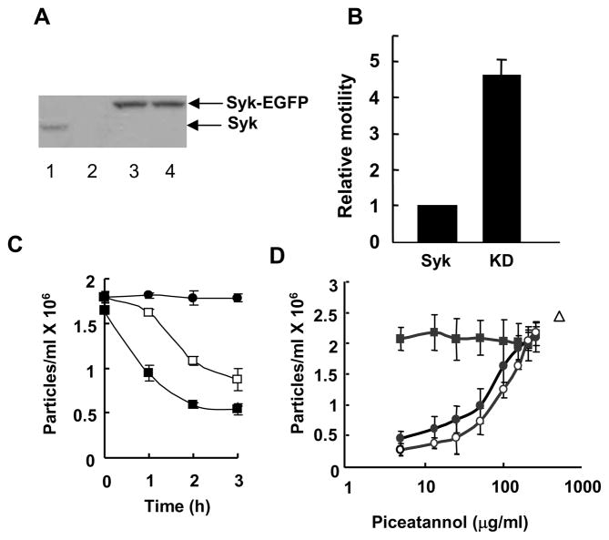
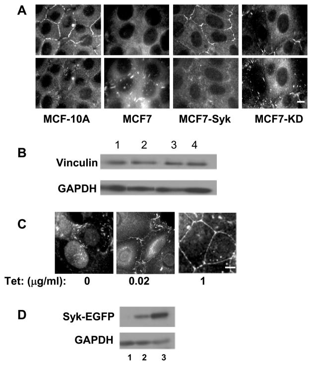
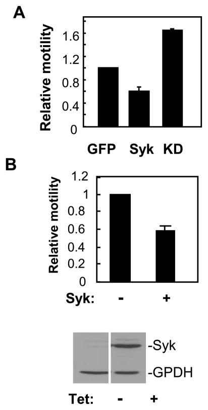
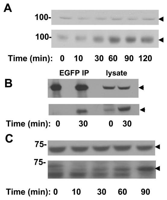
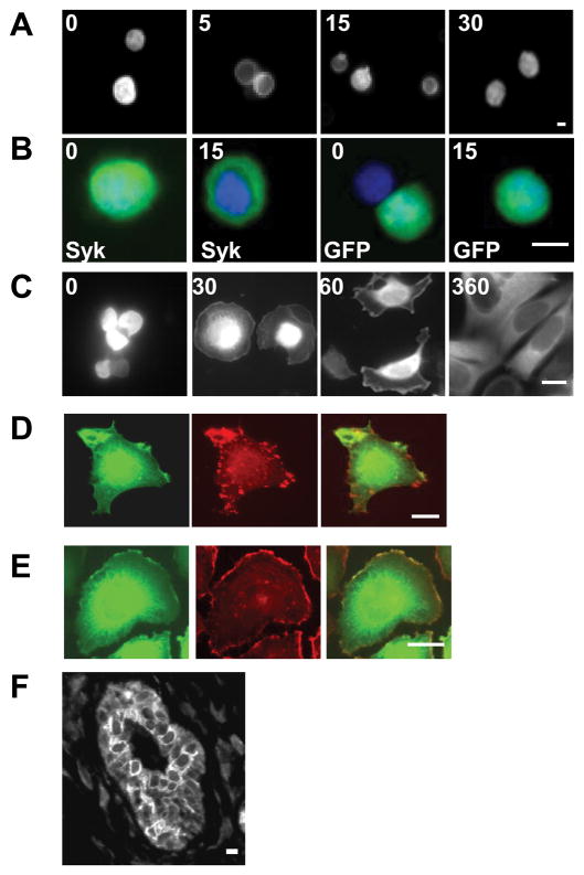
The expression of the Syk protein tyrosine kinase in breast cancer cells is inversely correlated with invasive growth and metastasis. The expression of Syk inhibits cell motility while supporting the formation of cell clusters by enhancing cell-cell contacts and promoting the redistribution of the adhesion proteins cortactin and vinculin to these contacts. Syk associates physically with cortactin and catalyzes its phosphorylation on tyrosine. The clustering of integrins leads to the phosphorylation of Syk and of numerous cellular proteins in a manner dependent on the activity of the kinase and on the presence of tyrosine 342 located in the linker B region. The ability of Syk to participate in integrin-mediated protein tyrosine phosphorylation correlates well with its ability to inhibit cell motility.


Figures

Figure 1
Figure 1
Syk alters cell aggregation and motility. A, lysates from MCF10A (lane1), Syk-deficient MCF7 (lane 2) or MCF7 cells stably expressing Syk-EGFP (lane 3) or Syk-EGFP(K396R) (lane 4) were analyzed by Western blotting with an antibody against Syk (N19). Arrows mark the migration positions of the Syk-EGFP and Syk-EGFP(K396R) fusion proteins (upper arrow) and endogenous Syk (lower arrow). B, MCF7 cells stably expressing Syk-EGFP (Syk) or Syk-EGFP(K396R) (KD) were analyzed in the transwell migration assay. The cells at the lower surface of the filter were stained with DAPI and observed using fluorescence microscopy. Motility was normalized to that of cells expressing Syk-EGFP. The data indicate the mean and standard deviations of three independent experiments (P < 0.01). C, stable MCF7 cell lines expressing Syk-EGFP (■) or Syk-EGFP(K396R) (□) were allowed to aggregate for the indicated periods of time. MCF7 cells did not aggregate in the presence of EGTA (●). The number of particles formed in four individual samples was counted. The averages and standard deviations for three experiments are shown. D, MCF10A cells were allowed to form cell-cell contacts for a period of 0 (■), 1 (●) or 2 (○) h in the presence of increasing concentrations of piceatannol. The number of particles formed in 2 h in the absence of piceatannol, but in the presence of EGTA is indicated (△). The number of particles is defined as a single cell or a single cluster of cells. The data indicate the mean and standard deviations of three independent experiments.
Figure 2
Figure 2
The expression of Syk causes a redistribution of vinculin. A, MCF10A, MCF7, and MCF7 cells expressing Syk-EGFP (MCF7-Syk) or Syk-EGFP(K396R) (MCF7-KD) were grown on glass coverslips, fixed, stained for vinculin and examined by fluorescence microscopy. Two focal planes including cell-cell contacts (upper panels) or focal adhesions (lower panels) are shown. B, Western blotting analysis of vinculin and glyceraldehyde-3-phosphate dehydrogenase (GAPDH) in lysates from MCF10A (lane 1), MCF7 (lane 2), MCF7-Syk (lane 3) and MCF7-KD (lane 4) cells. The images are derived from the same gel and blot, but, with the exception of lanes 3 and 4, are from nonadjacent lanes. C, Tet-response MCF7 cells were untreated (0) or treated with 20 ng/ml (0.02) or 1 μg/ml (1) tetracycline to induce expression of Syk-EGFP. Cells grown on untreated coverslips were fixed, stained for vinculin and examined by fluorescence microscopy. D, Western blotting analysis of Syk-EGFP and GAPDH in lysates of Tet-responsive MCF7 cells that were untreated (lane 1) or treated with 20 ng/ml (lane 2) or 1 μg/ml (land 3) tetracycline to induce expression of Syk-EGFP. Bars = 10μm.
Figure 3
Figure 3
Partial localization of Syk to cell-cell contacts. A, Tet-inducible MCF7 cells plated on glass coverslips were treated with tetracycline to induce Syk-EGFP and examined by fluorescence microscopy. B, MCF10A cells transiently expressing Syk-EGFP were treated with EGTA for 20 min, washed and placed in calcium-containing medium for 0, 20 or 45 min. Cells were fixed, permeabilized and stained for E-cadherin using DECMA-1 and a fluorescently tagged secondary antibody. Cells were examined for localization of Syk-EGFP (green) and E-cadherin (red) by fluorescence microscopy. Images were overlayed to produce the pictures in the bottom row of panels. C, MCF7 cells expressing Syk-EGFP(K396R) were fixed and examined by fluorescence microscopy. The arrowhead illustrates the localization of a subset of Syk-EGFP(K396R) to the plasma membrane. D, Syk-EGFP was transiently expressed in Ras-transformed MCF10A cells, which were fixed and examined by fluorescence microscopy. The arrowhead illustrates the weak localization of a subset of Syk-EGFP to the plasma membrane. E, Western blotting of lysates from Ras-MCF10A cells (lanes 1 and 2) or MCF10A cells (lane 3) with an anti-Syk antibody. Bars = 10 μm.
Figure 4
Figure 4
Syk interacts with cortactin. A, MCF7 cells or MCF7 cells expressing Syk-EGFP were grown on glass coverslips, fixed, stained for cortactin and examined by fluorescence microscopy. B, Lysates (lysate) or anti-cortactin immune complexes (cortactin IP) prepared from MCF7 (7), or MCF7 cells expressing Syk-EGFP (Syk) or Syk-EGFP(K396R) (KD) were analyzed by Western blotting using anti-cortactin or anti-Syk antibodies. C, lysates from MCF10A cells were adsorbed to protein G plus-agarose beads (Ctrl IP) or protein G plus-agarose beads containing immobilized anti-cortactin antibodies (Cort IP). Proteins in the lysate (lysate) or bound to the beads were immunoblotted using antibodies against cortactin (upper panel), Syk (middle panel) or E-cadherin (lower panel). D, lysates from MCF10A cells were adsorbed to protein G plus-agarose beads containing immobilized anti-Syk antibodies (Syk IP) or to protein G plus-agarose beads (Ctrl IP). Proteins in the lysate (lysate) or bound to the beads were immunoblotted using antibodies against Syk (upper panel) or cortactin (lower panel). E, GST or GST-Syk was incubated with a cortactin immune-complex isolated from MCF10A cells in a kinase reaction buffer containing [γ-32P]ATP. Protein G beads without antibody were used as a control. Samples were resolved by SDS-PAGE, transferred to a PVDF membrane and detected by autoradiography (32P) or Western blotting (WB) with anti-cortactin antibodies. F, lysates from MCF10A cells transiently transfected to express EGFP (lane 1) or Syk-EGFP (lane 2) were analyzed by Western blotting with anti-Syk antibodies (left panel). Confluent cultures of each were treated with EGTA to disrupt cell-cell contacts and then calcium was restored for a period of 25 min. Anti-cortactin immune complexes isolated from cell lysates were analyzed by Western blotting using anti-cortactin or anti-phosphotyrosine (pY) antibodies.
Figure 5
Figure 5
Syk inhibits MDA-MB-231 cell migration. A, MDA-MB-231 cells were transfected with expression plasmids for EGFP (GFP), Syk-EGFP (Syk) or Syk-EGFP(K396R) (KD) and analyzed in the transwell motility assay. The ratio of the number of green to blue cells at the lower surface of the filter was compared to that of initial cell population. This number from each sample was further normalized to values obtained for the migration of cells expressing EGFP alone. The data illustrate the mean and standard deviations of three independent experiments. P < 0.01. B, Tet-responsive MDA-MB-231 cells were left untreated (−) or were treated (+) with tetracycline and individually plated onto a transwell insert for the migration assay. Migrated cells at the lower surface of the two samples were counted and then normalized to the number of Syk-deficient (−) migrating cells. The data indicate the mean and standard deviations of three independent experiments. P < 0.01. The expression of Syk-EGFP and GADPH in Tet-treated (+) or untreated (−) cells was determined by Western blotting (lower panel).
Figure 6
Figure 6
Syk is activated by integrin-crosslinking in MDA-MB-231 cells. A, after being transiently transfected to express Syk-EGFP (Syk) or Syk-EGFP(K396R) (KD-Syk), MDA-MB-231 cells were removed and plated onto fibronectin-coated coverslips. After different time points (0, 10, 30, and 60 min), cell lysates were collected and analyzed by Western blotting with antiphosphotyrosine antibodies. The migration positions of Syk-EGFP or Syk-EGFP(K396R) are indicated by the arrowheads. B, integrins were crosslinked for the indicated times (in minutes) with anti-β1 integrin antibodies on MDA-MB-231 cells in suspension that were transiently expressing Syk-EGFP (Syk), Syk-EGFP(Y317F/Y342F/Y346F) (F3), EGFP (EGFP) or Syk-EGFP(K396R) (KD-Syk). Cell lysates were analyzed by Western blotting with antibodies against phosphotyrosine (pY), Syk or GAPDH. The migration positions of Syk-EGFP or Syk-EGFP mutants are indicated by the closed arrowheads and that of GAPDH by the open arrowheads. C, integrins were crosslinked for the indicated times (in minutes) with anti-β1 integrin antibodies on MDA-MB-231 cells in suspension that were transiently expressing Syk-EGFP(Y317F) (Y317F), Syk-EGFP(Y342F/Y346F) (Y342F/Y346F), Syk-EGFP (Syk), Syk-EGFP(Y342F) (Y342F) or Syk-EGFP(Y346F) (Y346F). Cell lysates were analyzed by Western blotting with antibodies against phosphotyrosine (pY), Syk or GAPDH. The migration positions of Syk-EGFP or Syk-EGFP mutants are indicated by the closed arrowheads and that of GAPDH by the open arrowheads. D, The ability of MDA-MB-231 cells transfected with expression plasmids for Syk-EGFP (Syk), Syk-EGFP(K396R) (KD-Syk), Syk-EGFP(Y317F/Y342F/Y346F) (F3), Syk-EGFP(Y317F) (Y317F), Syk-EGFP(Y342F/Y346F) (F2), Syk-EGFP(Y342F) (Y342F) or Syk-EGFP(Y346F) (Y346F) to migrate through the pores of a transwell filter was measured. Motility was normalized to that of cells expressing Syk-EGFP. The data shown represent the mean and standard deviations of three independent experiments. * P < 0.05.
Figure 7
Figure 7
Syk is phosphorylated following integrin-crosslinking. A, integrins on MCF7 cells expressing Syk-EGFP in suspension were crosslinked with anti-β1 integrin antibodies for the times indicated. Cell lysates were analyzed by Western blotting with antibodies against Syk (upper panel) or phosphotyrosine (lower panel). The migration position of Syk-EGFP is indicated by the arrowheads. B, lysate and anti-GFP immune complexes from MCF7 cells expressing Syk-EGFP and stimulated in suspension with anti-β1 integrin antibodies for the indicated times were analyzed by Western blotting with antibodies against Syk (upper panel) or phosphotyrosine (lower panel). The migration position of Syk-EGFP is indicated by the arrowheads. C, integrins on MCF10A cells in suspension were crosslinked with anti-β1 integrin antibodies for the times indicated. Cell lysates were analyzed by Western blotting with antibodies against Syk (upper panel) or phosphotyrosine (lower panel). The migration position of Syk is indicated by the arrowheads.
Figure 8
Figure 8
Integrin ligation alters the subcellular localization of Syk. A, MCF7 cells transfected with a Syk-EGFP-expression plasmid were stimulated by crosslinking β1 integrins for 0, 5, 15 or 30 min, fixed and examined by fluorescence microscopy. B, MCF7 cells transfected with plasmids expressing Syk-EGFP (Syk) or EGFP (GFP) were treated with β1 integrin antibodies for 0 or 15 min, fixed, stained with DAPI and examined by fluorescence microscopy. Merged images illustrating the location of Syk-EGFP or EGFP (green) and DAPI-stained nuclei (blue) are shown. C, Tet-inducible MCF7 cells treated with tetracycline to induce Syk-EGFP were plated on fibronectin-coated coverslips for the times indicated (in minutes) and then fixed and examined by fluorescence microscopy. D and E, MCF7 cells transfected with a Syk-EGFP-expression vector were plated on coverslips coated with fibronectin, allowed to spread for 60 min and then fixed, permeabilized and stained with monoclonal antibodies against vinculin (C) or with rhodamine phalloidin (D). The localization of Syk-EGFP (green, D, E), endogenous vinculin (red, D), and F-actin (red, E) were visualized with fluorescence microscopy. F, a human mammary tissue slice was stained with anti-Syk antibodies for immunohistochemical analysis. Bars = 10 μm.

References

    1. Turner M, Schweighoffer E, Colucci F, Di Santo JP, Tybulewicz VLJ. Tyrosine kinase SYK: essential functions for immunoreceptor signalling. Immunol Today. 2000;21:148–54. - PubMed
    1. Yanagi S, Inatome R, Takano T, Yamamura H. Syk expression and novel function in a wide variety of tissues. Biochem Biophys Res Commun. 2001;288:495–8. - PubMed
    1. Coopman PJ, Do MT, Barth M, et al. The Syk tyrosine kinase suppresses malignant growth of human breast cancer cells. Nature. 2000;406:742–7. - PubMed
    1. Yuan Y, Mendez R, Sahin A, Dai JL. Hypermethylation leads to silencing of the SYK gene in human breast cancer. Cancer Res. 2001;61:5558–61. - PubMed
    1. Moroni M, Soldatenkov V, Zhang L, et al. Progressive loss of Syk and abnormal proliferation in breast cancer cells. Cancer Res. 2004;64:7346–54. - PubMed

Publication types

MeSH terms