. 2009 Jul;297(1):E194-201.
doi: 10.1152/ajpendo.91041.2008. Epub 2009 May 12.

Diabetes increases brain damage caused by severe hypoglycemia

Affiliations

Diabetes increases brain damage caused by severe hypoglycemia

Adam J Bree et al. Am J Physiol Endocrinol Metab. 2009 Jul.

Abstract

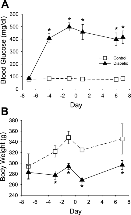
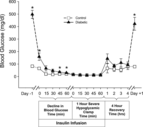
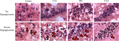
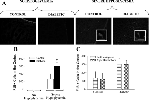
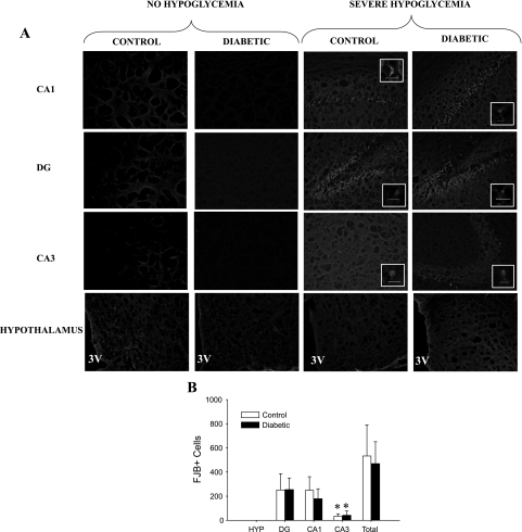
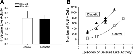
Insulin-induced severe hypoglycemia causes brain damage. The hypothesis to be tested was that diabetes portends to more extensive brain tissue damage following an episode of severe hypoglycemia. Nine-week-old male streptozotocin-diabetic (DIAB; n = 10) or vehicle-injected control (CONT; n = 7) Sprague-Dawley rats were subjected to hyperinsulinemic (0.2 U.kg(-1).min(-1)) severe hypoglycemic (10-15 mg/dl) clamps while awake and unrestrained. Groups were precisely matched for depth and duration (1 h) of severe hypoglycemia (CONT 11 +/- 0.5 and DIAB 12 +/- 0.2 mg/dl, P = not significant). During severe hypoglycemia, an equal number of episodes of seizure-like activity were noted in both groups. One week later, histological analysis demonstrated extensive neuronal damage in regions of the hippocampus, especially in the dentate gyrus and CA1 regions and less so in the CA3 region (P < 0.05), although total hippocampal damage was not different between groups. However, in the cortex, DIAB rats had significantly (2.3-fold) more dead neurons than CONT rats (P < 0.05). There was a strong correlation between neuronal damage and the occurrence of seizure-like activity (r(2) > 0.9). Separate studies conducted in groups of diabetic (n = 5) and nondiabetic (n = 5) rats not exposed to severe hypoglycemia showed no brain damage. In summary, under the conditions studied, severe hypoglycemia causes brain damage in the cortex and regions within the hippocampus, and the extent of damage is closely correlated to the presence of seizure-like activity in nonanesthetized rats. It is concluded that, in response to insulin-induced severe hypoglycemia, diabetes uniquely increases the vulnerability of specific brain areas to neuronal damage.


Figures

Fig. 1.
Fig. 1.
Effect of streptozotocin (STZ) on resting blood glucose and body weight. A: following STZ treatment, diabetic rats (n = 9; ▴) had markedly elevated blood glucose levels compared with buffer-treated control rats (n = 6; □). An episode of severe hypoglycemia was induced on day 0. B: diabetic rats failed to gain weight throughout the experiment and had significantly lower body weights than control rats throughout the experiment. *P < 0.05.
Fig. 2.
Fig. 2.
Blood glucose of rats before, during, and after severe hypoglycemia. Blood glucose levels of the control (n = 6; □) and diabetic (n = 9; ▴) rats are shown during the hyperinsulinemic severe hypoglycemic clamp as well as 1 day prior to and 1 day after the clamp. On the day of the clamp, insulin was infused intravenously at a constant rate (0.2 U·kg−1·min−1) to lower blood glucose. Once the blood glucose was ≤15 mg/dl, the clock was reset and glucose was infused to maintain a level of severe hypoglycemia between 10 and 15 mg/dl for 1 h. Following the 1 h of severe hypoglycemia, the insulin infusion was stopped and the rate of glucose infusion increased to restore euglycemia for 4 h. *P < 0.05.
Fig. 3.
Fig. 3.
Neuronal damage as assessed by hematoxylin and eosinophilic staining. Compared with rats that did not undergo hypoglycemia, neuronal damage induced following 1 h of severe hypoglycemia was evidenced by hematoxylin and eosinophilic staining. The rats that underwent severe hypoglycemia show brain cells characterized by condensed shrunken morphology and pyknotic nuclei (red arrows) with eosinophilic staining compared with the nuclei of rats that did not undergo hypoglycemia. This brain damage morphology is most evident in the cortex and specific regions of the hippocampus [CA1 and dentate gyrus (DG)]. ×400 Magnification with the scale bar indicating 50 microns.
Fig. 4.
Fig. 4.
Neuronal damage in the cortex. A: Fluoro-Jade B-positive (FJB+) staining in representative images taken at 3.1 mm posterior to Bregma in the lateral ectorhinal cortex superior to the rhinal fissure and viewed at ×100 magnification. A, insets: FJB+ cells viewed at ×400 magnification, with the bar indicating 100 microns. The 4 groups shown are nondiabetic control and STZ-diabetic rats that did not undergo hypoglycemia as well as nondiabetic control rats and STZ-diabetic rats that were subjected to severe hypoglycemia. B: there were no cortical FJB+ cells seen in rats that did not undergo hypoglycemia, whereas, in response to severe hypoglycemia, STZ-induced diabetes caused a 2.3-fold increase in the number of cortical FJB+ cells per brain section compared with nondiabetic controls. *P < 0.05. C: there was no difference in the amount of FJB+ cells in the left (bars with horizontal lines) vs. the right (cross-hatched bars) hemisphere of the brain in either control (n = 6) or diabetic (n = 9) rats.
Fig. 5.
Fig. 5.
Hippocampal and hypothalamic neuronal damage. A: FJB+ staining in the CA1, DG, and CA3 regions of the hippocampus and the hypothalamus viewed at ×100 magnification. A, insets: FJB+ cells viewed at ×400 magnification, with the bars indicating 100 microns. The pictures of the hypothalamus (HYP) were taken lateral to the 3rd ventricle (3V). The 4 groups shown are nondiabetic control and STZ-diabetic rats that did not undergo hypoglycemia, nondiabetic control rats that were subjected to severe hypoglycemia, and STZ-diabetic rats that were subjected to severe hypoglycemia. B: in response to severe hypoglycemia, both control (n = 5) and diabetic (n = 8) rats demonstrated no neuronal damage in the hypothalamus, but hippocampal neuronal damage was most evident in the DG and CA1 regions, with significantly less damage in the CA3 region. *P < 0.05.
Fig. 6.
Fig. 6.
A: correlation between seizure-like activity and neuronal damage. Both groups experienced the same number of seizure-like events (control 2.5 ± 0.7, diabetic 2.4 ± 0.4, P = NS). B: the amount of damage incurred in the cortex strongly correlates with the amount of seizure-like activity occurring during the 1 h of severe hypoglycemia [control r2 = 0.996 (□), n = 6; diabetic r2 = 0.945 (▴), n = 9].

References

    1. Alkire MT, Pomfrett CJ, Haier RJ, Gianzero MV, Chan CM, Jacobsen BP, Fallon JH. Functional brain imaging during anesthesia in humans: effects of halothane on global and regional cerebral glucose metabolism. Anesthesiology 90: 701–709, 1999. - PubMed
    1. Auer RN Hypoglycemic brain damage. Metab Brain Dis 19: 169–175, 2004. - PubMed
    1. Auer RN, Hugh J, Cosgrove E, Curry B. Neuropathologic findings in three cases of profound hypoglycemia. Clin Neuropathol 8: 63–68, 1989. - PubMed
    1. Austin EJ, Deary IJ. Effects of repeated hypoglycemia on cognitive function: a psychometrically validated reanalysis of the Diabetes Control and Complications Trial data. Diabetes Care 22: 1273–1277, 1999. - PubMed
    1. Baird TA, Parsons MW, Phanh T, Butcher KS, Desmond PM, Tress BM, Colman PG, Chambers BR, Davis SM. Persistent poststroke hyperglycemia is independently associated with infarct expansion and worse clinical outcome. Stroke 34: 2208–2214, 2003. - PubMed

Publication types