. 2009 May 15;69(10):4192-201.
doi: 10.1158/0008-5472.CAN-09-0042. Epub 2009 May 12.

Loss of Phosphatase and Tensin homologue deleted on chromosome 10 engages ErbB3 and insulin-like growth factor-I receptor signaling to promote antiestrogen resistance in breast cancer

Affiliations

Loss of Phosphatase and Tensin homologue deleted on chromosome 10 engages ErbB3 and insulin-like growth factor-I receptor signaling to promote antiestrogen resistance in breast cancer

Todd W Miller et al. Cancer Res. .

Abstract

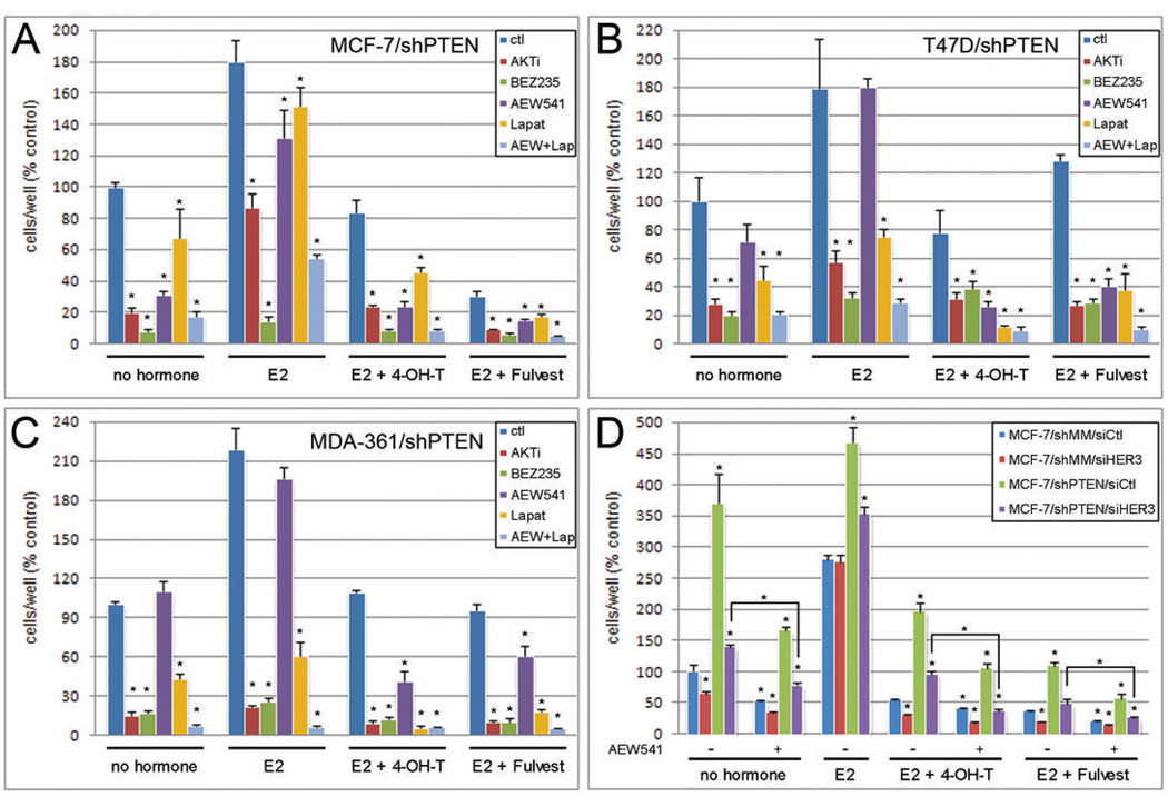
Knockdown of the tumor suppressor phosphatase Phosphatase and tensin homologue deleted on chromosome 10 (PTEN) with shRNA in three estrogen receptor (ER)-positive breast cancer cell lines resulted in increased phosphatidylinositol-3 kinase (PI3K) and AKT activities, resistance to tamoxifen and fulvestrant, and hormone-independent growth. PTEN knockdown induced the up-regulation of ER transcriptional activity in MCF-7 cells but decreased ER protein levels and transcriptional activity in T47D and MDA-361 cells. Tamoxifen and fulvestrant treatment inhibited estradiol-induced ER transcriptional activity in all shPTEN cell lines but did not abrogate the increased cell proliferation induced by PTEN knockdown. PTEN knockdown increased basal and ligand-induced activation of the insulin-like growth factor-I (IGF-I) and ErbB3 receptor tyrosine kinases, and prolonged the association of the p85 PI3K subunit with the IGF-I receptor (IGF-IR) effector insulin receptor substrate-1 and with ErbB3, implicating PTEN in the modulation of signaling upstream of PI3K. Consistent with these data, PTEN levels inversely correlated with levels of tyrosine-phosphorylated IGF-IR in tissue lysate arrays of primary breast cancers. Inhibition of IGF-IR and/or ErbB2-mediated activation of ErbB3 with tyrosine kinase inhibitors restored hormone dependence and the growth inhibitory effect of tamoxifen and fulvestrant on shPTEN cells, suggesting that cotargeting both ER and receptor tyrosine kinase pathways holds promise for the treatment of patients with ER+, PTEN-deficient breast cancers.


Figures

Fig. 1
Fig. 1. PTEN loss increases PIP3 and P-AKT, and alters ER and PR levels
A) Cells were treated with medium containing DCC-FBS (MCF-7: 2%; T47D, MDA-361: 0.5%) × 24 hrs, and lysates were analyzed by immunoblotting with the indicated antibodies. PR was not detected in MDA-361 cells. B) MCF-7 lines were metabolically labeled with 32P-orthophosphatase in 10% dialyzed FBS × 16 hrs. Lipids were extracted, resolved by thin-layer chromatography, and 32P-PIP species were detected by autoradiography (arrows). Origin of spotting is indicated. Cell lysates were also used for immunoblotting to confirm PTEN status. Fold-changes in PR isoforms (A) and PIP3 (B) normalized to actin were determined by densitometry analysis (bar graphs).
Fig. 2
Fig. 2. PTEN loss increases hormone-independent growth and antiestrogen resistance
A) Cell proliferation assays. MCF-7 (i, iv), T47D (ii), and MDA-361 (iii) lines were treated with medium containing DCC-FBS (MCF-7: 2%; T47D, MDA-361: 0.5%) with the indicated compounds (E2-17-β-estradiol; 4-OH-T- 4-hydroxy-tamoxifen; Fulvest- Fulvestrant; Lz- letrozole). Media and drugs were refreshed every 2–3 days. Adherent cells were counted after 5–8 days. B) ER transcriptional reporter assays. MCF-7 (i, iv), T47D (ii), and MDA-361 (iii) lines were transfected with luciferase reporter plasmids. Cells were treated as in (A), and luciferase activities were measured after 16–20 hrs. RLU- relative light units (firefly/Renilla). All data are presented as % untreated shMM control, mean of triplicates +/− SD. * p<0.05 by t-test comparing shPTEN to shMM under each condition.
Fig. 3
Fig. 3. PTEN loss activates RTKs upstream of PI3K
A) Lysates from MCF-7 (i), T47D (ii), and MDA-361 (iii) lines treated with medium containing DCC-FBS (MCF-7: 2%; T47D, MDA-361: 0.5%) overnight were used to probe phospho-RTK arrays. Antibodies against 42 RTKs are spotted in duplicate on a membrane. Membranes are incubated with cell lysates followed by probing with a P-tyr antibody. A positive signal is indicative of receptor phosphorylation. Blots from each pair of lines are exposure-matched. Tyr-phosphorylated RTKs are labeled as: 1-EGFR; 2-HER2; 3-HER3; 4-InsRβ; 5-IGF-IRβ; 6-ErbB4; 7-ROR2; 8-EphA1. Positive controls are spotted at corners. B) Lysates from MCF-7 (i), T47D (ii), and MDA-361 (iii) lines treated as in (A) overnight +/− the indicated kinase inhibitors were used for immunoblotting with the indicated antibodies. Short and long exposures (exp) are shown in (iii). C) Tumor lysates from 383 hormone receptor-positive breast cancers were analyzed by RPPA to quantify relative levels of PTEN, P-IGF-IRβY1135/1136, and IGF-IRβ. Shown is a scatterplot of PTEN vs. ratio of P-IGF-IRβ/IGF-IRβ (indicative of the fraction of activated IGF-IRβ). This relationship was analyzed using two-tailed t-test and Pearson correlation.
Fig. 4
Fig. 4. PTEN loss increases PI3K activation and sensitivity to RTK ligands
A) p85 was immunoprecipitated from cell lysates of MCF-7 (i), T47D (ii), and MDA-361 (iii) lines treated overnight +/− 10% DCC-FBS. Short and long exposures (exp) are shown in (ii). B) Immunoblotting with the indicated antibodies of i) lysates from MCF-7 cells serum-starved overnight, then treated with IGF-I (0–100 ng/mL × 15 min.); ii) lysates from T47D and MDA-361 lines treated overnight +/− 10% DCC-FBS; iii) lysates from T47D cells serum-starved overnight, then treated with heregulin-β1 (20 ng/mL × 0–12 min.).
Fig. 5
Fig. 5. PTEN loss prolongs IGF-IR and HER3 signaling, and increases E2-induced non-genomic signaling via IGF-IR
A) p85 was immunoprecipitated from lysates of MCF-7 cells that had been pretreated overnight with serum-free medium +/− AEW541 (1 µM), then stimulated +/− 100 ng/mL IGF-I +/− AEW541 × 5, 60, or 180 min. Arrowhead indicates P-IGF-IRβ. B) Lysates from MCF-7 cells pretreated as in (A) +/− AEW541 (1 µM) or lapatinib (1 µM), then stimulated +/− IGF-I (100 ng/mL) +/− inhibitors × 15 min. C) p85 was immunoprecipitated from lysates of T47D cells treated with 0.5% DCC-FBS +/− lapatinib [1 µM × 15, 30, 60, 120, or 180 min., or overnight (o/n)]. Short and long exposures (exp) for P-HER3 are shown. D) p85 was immunoprecipitated from lysates of MCF-7 cells pretreated overnight with 10% DCC-FBS +/− 1 µM 4-OH-T, 1 µM fulvestrant, 1 µM AEW541, or 1 µM lapatinib, and then stimulated +/− 1 nM E2 +/− inhibitors × 20 min. Arrowhead indicates tyr-phosphorylated IRS-1 (≈150 kDa). All immunoprecipitates and cell lysates were analyzed by immunoblotting with the indicated antibodies.
Fig. 6
Fig. 6. Combined inhibition of IGF-IR and EGFR/HER2 synergizes with 4-OH-T, fulvestrant, and hormone-deprivation to block cell proliferation
A) MCF-7/shPTEN, B) T47D/shPTEN, and C) MDA-361/shPTEN cells were treated with medium containing DCC-FBS (MCF-7: 2%; T47D, MDA-361: 0.5%) as indicated [0.1 nM E2 (MCF-7), 1 nM E2 (T47D, MDA-361), 1 µM 4-OH-T, 1 µM fulvestrant, 1 µM AKTi, 200 nM BEZ235, 1 µM AEW541, 1 µM lapatinib]. Media and drugs were refreshed every 2–3 days. Adherent cells were counted after 5–8 days. Data are presented as % untreated shMM control, mean of triplicates +/− SD. * p<0.05 by t-test comparing kinase inhibitor-treated cells to control (ctl) cells within each group. D) MCF-7/shPTEN and /shMM cells transfected with siRNA against HER3 or control (siCtl) were treated and analyzed as in (A). * p<0.05 by t-test compared to untreated shMM/siCtl within each group.

References

    1. Li J, Yen C, Liaw D, et al. PTEN, a putative protein tyrosine phosphatase gene mutated in human brain, breast, and prostate cancer. Science. 1997;275:1943–1947. - PubMed
    1. Saal LH, Holm K, Maurer M, et al. PIK3CA mutations correlate with hormone receptors, node metastasis, and ERBB2, and are mutually exclusive with PTEN loss in human breast carcinoma. Cancer Res. 2005;65:2554–2559. - PubMed
    1. Depowski PL, Rosenthal SI, Ross JS. Loss of expression of the PTEN gene protein product is associated with poor outcome in breast cancer. Mod Pathol. 2001;14:672–676. - PubMed
    1. Perren A, Weng LP, Boag AH, et al. Immunohistochemical evidence of loss of PTEN expression in primary ductal adenocarcinomas of the breast. Am J Pathol. 1999;155:1253–1260. - PMC - PubMed
    1. Tamura M, Gu J, Matsumoto K, Aota S, Parsons R, Yamada KM. Inhibition of cell migration, spreading, and focal adhesions by tumor suppressor PTEN. Science. 1998;280:1614–1617. - PubMed

Publication types